https://doi.org/10.36253/caryologia-3030
Received: September 20, 2024; Accepted: June 21, 2025; Published: October 1, 2025
Cytogenetic diagnosis of patients with suspected premature ovarian failure in Manaus, Brazil
1 Laboratório de Citogenética Humana, Universidade do Estado do Amazonas, Manaus, Brasil
2 Instituto de Saúde e Biotecnologia, Universidade Federal do Amazonas, Coari, Brasil
3 Departamento de Ginecologia, Universidade do Estado do Amazonas, Manaus, Brasil
*Corresponding author. Email: cfantin@uea.edu.br
Abstract. Premature ovarian failure (POF) is a clinical syndrome that is characterized by loss of ovarian function in women of childbearing age and generally occurs before the age of 40. Genetic causes account for about 20 to 25% of cases of POF. However, in many cases, the origin of the condition remains idiopathic. The objective of this study was to perform cytogenetic research in a group of patients affected by POF in order to identify the type and frequency of chromosomal alterations. Fifteen patients were referred to the Human Cytogenetics Laboratory of the Amazonas State University (UEA) by gynecology specialists from two public health institutions in Manaus, Amazonas, Brazil, for chromosomal analysis. The analysis was performed via peripheral blood lymphocyte culture using the GTG banding method. The karyotypes were assembled with the help of the GeneAll-HD® software and the results were interpreted according to the ISCN 2016 standards. Of the fifteen patients analyzed, nine (60%) had no chromosomal abnormalities, while six (40%) exhibited chromosomal abnormalities. Of the alterations identified, three patients (20%) presented numerical alterations of the X chromosome with mosaicism, two patients (13%) showed autosomal numerical alterations involving chromosomes 15 and 21, both with mosaicism, and one patient (7%) exhibited a structural alteration in the form of terminal deletion of the long arm of the X chromosome. The results obtained in this study have the potential to improve the accuracy of the diagnosis, assist in medical decisions, provide adequate prognoses and facilitate reproductive management through genetic counseling.
Keywords: Premature Ovarian Failure, G-band karyotype, chromosomal alterations, X chromosome, medical diagnosis.
Contents
Introduction
Premature ovarian failure (POF) is a clinical syndrome that is defined by the loss of ovarian function in women of childbearing age, generally before the age of 40. The main symptoms of POF are menstrual disorders such as amenorrhea or oligomenorrhea, as well as having high levels of gonadotropins and low levels of estradiol. These symptoms result in hypoestrogenic and hypergonadotropic clinical pictures (Goswami and Conway 2005; Eshre 2016; Chon 2021). The first reports of this syndrome were made in 1942 by Fuller Albright, who named the condition “primary ovarian failure” (Albright 1942).
The overall incidence of POF is estimated to be approximately 1% in women aged under 40 and 0.1% in women under 30 (Rahman and Panay 2021). In the mid and long term, this condition can cause an increase in cardiovascular diseases, a decrease in bone mineral density with a risk of osteoporosis, and a progressive decline in fertility with neurological effects, resulting in a general reduction in the woman’s life expectancy (Podfigurna-Stopa et al. 2016; Wesevich 2020).
The etiology of POF is highly heterogeneous and causes can be genetic, autoimmune, metabolic, infectious and iatrogenic (Jiao et al. 2012; Qin 2015). Genetic causes account for approximately 20 to 25% of patients with POF (Ayed et al. 2014; Qin 2015; Luo et al. 2023). The most common genetic cause of POF is alterations involving the X chromosome. These changes can be of the numerical type, such as monosomy X, trisomy X, mosaicism X, or structural changes, such as deletions X, X-autosome translocations and isochromosomes (Holland 2001; Baronchelli et al. 2011).
In the diagnosis of POF, a complete gynecological evaluation is carried out with physical, biochemical and imaging examinations, which mainly include the measurement of high gonadotropin levels and low estradiol levels in patients under the age of 40 years who have symptoms of oligomenorrhea or amenorrhea lasting at least 4 months. The specialist can ascertain this condition when examining women with menstrual disorders. In cases of secondary amenorrhea, it is necessary to exclude pregnancy by examining serum levels of beta-hCG. After confirming the diagnosis of POF through physical and biochemical examinations, due to its heterogeneity, it becomes necessary to request complementary examinations, such as cytogenetic analyses including karyotyping, in order to determine the etiology (Eshre 2016; Jankowska 2017).
Cytogenetic and molecular investigations of these alterations have allowed us to identify two critical regions in the long arm of the X chromosome, in Xq13-q21 and Xq26-27 (Powell et al. 1994; Sala et al. 1997). Although chromosomal alterations primarily involve the X chromosome, an increasing number of alterations involving autosomal chromosomes have been reported in the literature (Goswami and Conway 2005). Chromosomal abnormalities have long been recognized as a frequent cause of POF, with widely varying percentages reported in the literature in both primary and secondary amenorrhea, thus suggesting the need for cytogenetic analyses (Lakhal et al. 2010; Ceylaner et al. 2010; Kalantari et al. 2013; Ayed et al 2014). Nonetheless, in most cases, the etiopathogenesis of this condition still remains idiopathic (Rudnicka et al. 2018).
Given the strong impact of the disease on the quality of life of these women, it is important to highlight the value of cytogenetic investigations, including karyotyping, which can be requested for women affected by POF, in order to detect the presence of chromosomal alterations that may be associated with this condition (Di-Battista 2020). In Brazil, most studies involving the cytogenetic diagnosis in women affected with POF are concentrated in the southern and southeastern regions of the country and cytogenetic investigations related to POF in the state of Amazonas have not yet been carried out.
Therefore, the purpose of this study was to conduct cytogenetic analyses in patients affected by this condition. The results obtained have the potential to offer crucial information to the medical and scientific community of the northern region of Brazil. These findings not only fill a gap in current knowledge, but also have the potential to drive the development of more effective prevention and treatment strategies for POF.
Materials and methods
Patients
A prospective study was conducted in patients with suspected premature ovarian failure who were seen at two public health institutions in Manaus, Amazonas, Brazil: the department of Climacteric, Gynecology and Mastology in the Araújo Lima outpatient clinic at the Getúlio Vargas University Hospital (HUGV), and the department of Gynecology and Obstetrics at the Codajás Polyclinic.
The inclusion criteria for the study were as follows: patients treated by the Unified Health System (SUS) in Manaus, Amazonas, and affected by POF according to the guidelines of the European Society of Human Reproduction and Embryology ESHRE (Eshre 2016). Criteria for diagnosis of POF included: (I) primary or secondary oligo/amenorrhea for at least four consecutive months before the age of 40. (II) high levels of FSH >25 IU/mL and low levels of estradiol <20 pg/mL in the blood, (III) in two dosages more than 4 weeks apart. Patients with conditions known to induce POF, such as chemotherapy or radiotherapy and ovarian surgery, were excluded.
The study was approved by the Ethics Committee of the Universidade do Estado do Amazonas (UEA), under CAAE number 95704617.0.0000.5016. The participation of the patients was voluntary and an informed consent form was signed by all the participants. A total of 15 patients were referred to the Human Cytogenetics Laboratory of the Universidade do Estado do Amazonas, where 5 mL of peripheral blood was collected by venipuncture with a disposable sterile syringe for subsequent culture of lymphocytes.
Cytogenetic analysis
The karyotype analysis was performed using peripheral blood lymphocyte cultures following the methodology described by Moorhead et al. (1960), with modifications. The GTG-banding technique was performed according to Seabright (1973), with modifications. The resolution obtained was 400-500 bands per genome. The karyotype was determined using an optical microscope (Coleman® Trinocular N126T-Infinito Plano Led), with 30 metaphases per patient being analyzed and, in cases of suspected mosaicism, this number was increased to 50 metaphases. The metaphases were photographed using ScopeImage software (version 9.0) and karyotyped using GeneAll-HD software. The results obtained from the GTG-banding were interpreted according to the norms present in the International System for Human Cytogenetic Nomenclature ISCN (McGowan-Jordan 2016).
Statistical analysis
Statistical analyses were performed based on data obtained from patient records, which were compiled and analyzed using Microsoft Excel® software. The relative frequency of the following parameters were obtained: distribution of the results of the normal and altered karyotypes involved (autosomal or sexual); and were relative to the frequency of chromosomal analyses in POF patients.
Results
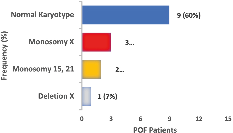
Between April 2022 and February 2023, chromosomal analyses were conducted at the Laboratory of Human Cytogenetics (UEA) in 15 patients suspected of premature ovarian failure (POF). The results revealed that nine of these patients (60%) presented a normal karyotype 46,XX, without any numerical or structural chromosomal alterations, representing the majority of the cases analyzed. However, in six patients (40%), numerical or structural chromosomal alterations involving autosomal chromosomes and the X chromosome were identified (Table 1).
| Nº | Age | Alterations | Chromosome | Karyotype |
|---|---|---|---|---|
| 001 | 19 | Numerical-Monosomy-Mosaic | Sexual | 45,X[4]/46,XX[46] |
| 002 | 36 | Numerical-Monosomy-Mosaic | Sexual | 45,X[3]/46,XX[47] |
| 003 | 37 | Numerical-Monosomy-Mosaic | Sexual | 45,X[4]/46,XX[46] |
| 004 | 34 | Numerical-Monosomy-Mosaic | Autosomal | 45,XX,-15[6]/46,XX[44] |
| 005 | 34 | Numerical-Monosomy-Mosaic | Autosomal | 45,XX,-21[5]/46,XX[45] |
| 006 | 22 | Structural-Deletion-Non-mosaic | Sexual | 46,X,del(X)(q22-24;qter) |
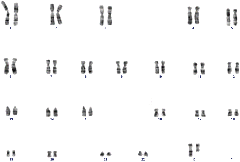
The prevalence of chromosomal alterations found in patients with POF was as follows: 20% of the patients presented numerical alterations of the monosomy type of the X chromosome with mosaicism, while 13% demonstrated numerical alterations in autosomal chromosomes, including pairs 15 and 21, both with mosaicism (Figure 1). In addition, 7% of patients exhibited a structural alteration characterized by a terminal deletion in the long arm of the X chromosome (Figure 2).
Discussion
According to Chen et al. (2023), the cytogenetic analysis of blood lymphocytes, using the karyotype, is an important tool in the detection of chromosomal alterations, both numerical and structural, and plays a crucial role in understanding the underlying genetic causes of POF.
This work is the first cytogenetic study using peripheral blood of a population of women affected by POF in the northern region of Brazil, since most studies involving cytogenetic diagnosis in women with POF are performed in the southern and southeastern regions of the country. In our study, six patients with POF were identified as having numerical or structural chromosomal alterations involving both autosomal chromosomes and the X chromosome, with these alterations representing 40% of the total number of cases.
One study conducted in patients affected by POF who were submitted to karyotype examinations in a public community genetics service in the state of Rio Grande do Sul, Brazil, demonstrated that 41 (29%) of the 141 confirmed cases presented numerical and structural alterations related to the X chromosome (Rosa et al. 2008). In another retrospective study involving 43 women affected by POF, conducted in public genetic services of the southern region of the country, Besson et al. (2023) identified, mainly in the X chromosome, numerical and structural chromosomal alterations in 14 (32.6%) of the participating women.
Our results also resemble those of a Turkish cytogenetic study involving women affected by POF, which identified a prevalence of chromosomal alterations in 39 (52%) of the 75 participating patients (Ceylaner et al. 2010). On the other hand, in another Turkish cytogenetic study, a lower prevalence of numerical and structural chromosomal alterations involving both autosomal chromosomes and the X chromosome was identified in 44 (25%) of the 175 cases analyzed in women with POF (Geckinli et al. 2014). In an Egyptian cytogenetic study involving 30 women with POF, it was observed that seven cases (23.3%) had chromosomal alterations, including numerical and structural alterations in the X chromosome (Issa and Elhady 2022).
In a Chinese cytogenetic study, chromosomal alterations were present in 64 (12.1%) of the 531 cases of POF analyzed (Jiao et al. 2012). In addition, a study involving Tunisian women with POF showed that 10.8% (108) of the 1,000 patients submitted to karyotype analysis had chromosomal alterations, mainly numerical ones of the X chromosome (Lakhal et al. 2010). Significant differences in the percentage of chromosomal alterations identified in various studies involving POF may be associated with ethnicity (Luborsky 2003). Luborsky et al. (2003) conducted a multi-ethnic epidemiological study and identified variations in the occurrence of POF, with the largest differences observed in Caucasian, African American and Hispanic women. Thus, the prevalence of POF may present ethnical differences, as well as regional ones due to lifestyle and environmental factors, which still need to be more fully investigated (Ishizuka 2021).
In our study, we identified that the highest prevalence of chromosomal abnormalities was of X chromosome monosomy, with a low frequency of mosaicism in three patients, representing 20% of the cases analyzed. The result obtained here is in line with what was found in a study that investigated women with POF and identified a 21.9% prevalence of chromosomal alterations with the presence of low frequency mosaicism of the X chromosome in the cases analyzed (Gersak and Veble 2011).
The most common chromosomal alteration in a cytogenetic study involving 179 Iranian women was X-chromosome mosaicism, which was present in 27.77% of cases (Kalantari 2013). It is said in the literature that karyotypes 45,X, with or without mosaicism, in numerical alterations of the X chromosome, can manifest themselves clinically with symptoms of primary or secondary amenorrhea (Turkyilmaz 2022). X-chromosome mosaicism is usually associated with sexual development and abnormal reproductive performance in women, resulting in infertility, recurrent miscarriages and cases of POF (Gersak and Veble 2011).
The results confirm previous observations and emphasize the critical role of alterations involving the X chromosome, with low frequency mosaicism, as one of the possible etiologies of POF (Ceylaner et al. 2010). Two intact X chromosomes are essential for the maintenance of ovarian function, since many genes that are likely involved in ovarian function escape X inactivation and are necessary for normal ovarian function, development, and maintenance (Davison 1999; Zinn 2001). In addition, low-frequency mosaicism involving the X chromosome can influence the survival rate and accelerate the aging of ovarian cells through different mechanisms, including a decrease in the number of germ cells or acceleration of their postnatal destruction and early oocyte atresia (Neves et al. 2020; Issa and Elhady 2022).
Other numerical alterations of the monosomy type, involving autosomal chromosomes 15 and 21, were observed in our study. The alterations were found in two nulliparous patients (34-year-old monozygotic twin sisters), representing 13% of the cases. In both cases, a low-frequency mosaicism was observed. The literature describes that POF is related to familial occurrence in about 12% to 15% of cases (Van Kasteren et al. 1999; Ferrarini et al. 2013; Franić et al. 2016; Rudnicka et al. 2018). To date, in the literature, there have been no reports of patients with POF who presented the absence of autosomal chromosomes 15 or 21 in their karyotypes.
However, Hosseini et al. (2011), reported a case of a 27-year-old Iranian woman affected by POF who presented a balanced translocation between autosomal chromosomes 15 and 21. The literature describes the occurrence of partial deletion of 21q in a case of a woman affected by POF (Zeng 2019). Another case of POF was reported in a 36-year-old woman, in which cytogenetic analyses revealed the presence of a supernumerary marker, which was characterized by fluorescent in situ hybridization (FISH) and comparative genomic hybridization (array CGH). The marker was derived from chromosome 15 and contained only heterochromatic material (Bertini 2012). Another study identified a patient with POF who had an autosomal mosaicism involving trisomy 21 (Baronchelli et al. 2011).
Other autosomal chromosome-related changes have been identified in patients with POF; in a study conducted by Issa et al. (2022), reciprocal translocations between the X chromosome and chromosome 9 were found. In another study, conducted by Jiao et al. (2012), a Robertsonian translocation between chromosomes 13 and 14 was identified in women affected by POF.
Regarding the structural alterations identified in the present study, we found one patient (7%) who presented a terminal deletion in the long arm of an X chromosome in all the metaphases examined. Similar findings were observed in a cytogenetic study in women affected by POF in Egypt, which identified a terminal deletion in the long arm on one of the X chromosomes, in the absence of mosaicism in all the metaphases analyzed (Issa and Elhady 2022). The complete or partial absence of an X chromosome, as seen in Turner syndrome, leads to ovarian dysgenesis characterized by primary amenorrhea and phenotypic features such as short stature (Goswami and Conway 2005; Portnoi et al. 2006). However, terminal deletions of the chromosome Xq are among the most common cytogenetic alterations. Two critical regions associated with POF have been defined in previous studies on X chromosome rearrangements: Xq13-Xq21 and Xq23-Xq27; these regions have been characterized for ovarian development and function (Persani 2009; Beke et al. 2013; Barros 2020; Besson et al. 2023).
Previous studies have shown that deletion of both the short arm and the long arm of the X chromosome can result in primary or secondary amenorrhea (Sybert and McCauley 2004). These observations suggest that genes that are important for normal ovarian function are located on both arms of the X chromosome (Cordts 2010). The high frequency of chromosomal alterations found in the present study emphasizes the importance of routine cytogenetic (karyotyping) tests in the investigation of patients affected with POF, for diagnostic definition and adequate genetic counseling for patients and their families.
However, even in the face of normal karyotyping results, the possibility of genetic alterations cannot be ruled out, since possible mutations in different genes can cause POF. The most common single-gene cause that results in this condition is pre-mutation of the FMR1 gene, located on the long arm of the X chromosome. This mutation is based on increased expansion of CGG trinucleotide repeats from 55 to 199 in the untranslated region. Patients with this pre-mutation have an increased risk of developing POF (Jin 2012; Rudnicka et al. 2018).
Therefore, a more detailed investigation is necessary for these patients and should involve tests using molecular techniques for diagnostic confirmation.
Conclusion
POF continues to be a serious medical problem and significantly affects the patient’s life. In most cases, its etiopathology remains unexplained. The relationship between chromosomal alterations and some cases of POF is clearly demonstrated in the present study. The cytogenetic findings highlight the importance of chromosomal analysis via conventional cytogenetics in the investigation of this condition. In most cases, it was possible to identify both numerical (20%) and structural (7%) chromosomal alterations, mainly involving the X chromosome, including low frequency mosaicism, which is directly associated with this condition.
In addition, it was possible to detect numerical alterations of the monosomy type involving autosomal chromosomes (13%) referring to pairs 15 and 21. This information is fundamental in clinical practice for the correct diagnosis, adequate prognosis and reproductive management through genetic counseling. Such results aim to contribute to medical decision-making during its diagnosis and help direct a multidisciplinary therapeutic approach by specialists.
Early diagnosis of POF is extremely important for maintaining the physical and mental health of these patients, as it can provide etiological explanation, help in the prevention of bone and cardiovascular health, and guide fertility options. Thus, the diagnosis represents a fundamental milestone in promoting the quality of life of these patients. These findings are essential to the understanding of POF and may have significant implications in the diagnosis and management of this condition.
Acknowledgments
This work was supported by the Fundação de Amparo à Pesquisa do Estado do Amazonas (FAPEAM) and the Coordenacão de Aperfeiçoamento de Pessoal de Nível Superior (CAPES).
References
Albright F, Smith PH, Fraser R. 1942. A syndrome characterized by primary ovarian insufficiency and decreased stature: Report of 11 cases with a digression on hormonal control of axillary and pubic hair. Am J Med Sci. 204(5):625–648. https://doi.org/10.1097/00000441-194211000-00001.
Ayed W, Amouri A, Hammami W, Kilani O, Turki Z, Harzallah F, Bouayed-Abdelmoula N, Chemkhi I, Zhioua F, Slama CB, et al. 2014. Cytogenetic abnormalities in Tunisian women with premature ovarian failure. C R Biol. 337(12):691–694. https://doi.org/10.1016/j.crvi.2014.09.003.
Baronchelli S, Conconi D, Panzeri E, Bentivegna A, Redaelli S, Lissoni S, Saccheri F, Villa N, Crosti F, Sala E, et al. 2011. Cytogenetics of premature ovarian failure: an investigation on 269 affected women. J Biomed Biotechnol. 2011(1):370195. https://doi.org/10.1155/2011/370195.
Barros F, Carvalho F, Barros A, Dória S, et al. 2020. Premature ovarian insufficiency: clinical orientations for genetic testing and genetic counseling. Porto Biomed J. 5(3):e62. https://doi.org/10.1097/j.pbj.0000000000000062.
Beke A, Piko H, Haltrich I, Csomor J, Matolcsy A, Fekete G, Rigo J, Karcagi V, et al. 2013. Molecular cytogenetic analysis of Xq critical regions in premature ovarian failure. Mol Cytogenet. 6(1):62. https://doi.org/10.1186/1755-8166-6-62.
Bertini V, Viola D, Vitti P, Simi P, Valetto A, et al. 2012. An idic(15) associated with POF (premature ovarian failure): Molecular cytogenetic definition of a case and review of the literature. Gene. 503(1):123–125. https://doi.org/10.1016/j.gene.2012.04.071.
Besson M da R, Taiarol M dos S, Fernandes EB, Ghiorzi IB, Nunes MR, Zen PRG, Rosa RFM, et al. 2023. Chromosomal abnormalities detected by karyotyping among patients with secondary amenorrhea: a retrospective study. Sao Paulo Med J. 141(5). https://doi.org/10.1590/1516-3180.2022.0426.r1.14012023.
Geckinli, Toksoy, Sayar, Soylemez, Yesil, Aydın, Karaman, Devranoglu, et al. 2014. Prevalence of X-aneuploidies, X-structural abnormalities and 46,XY sex reversal in Turkish women with primary amenorrhea or premature ovarian insufficiency. Eur J Obstet Gynecol Reprod Biol. 182:211–215. https://doi.org/10.1016/j.ejogrb.2014.09.033.
Ceylaner G, Altinkaya SO, Mollamahmutoglu L, Ceylaner S, et al. 2010. Genetic abnormalities in Turkish women with premature ovarian failure. Int J Gynecol Amp Obstet. 110(2):122–124. https://doi.org/10.1016/j.ijgo.2010.03.023.
Chen M, Jiang H, Zhang C. 2023. Selected Genetic Factors Associated with Primary Ovarian Insufficiency. Int J Mol Sci. 24(5):4423. https://doi.org/10.3390/ijms24054423.
Chon SJ, Umair Z, Yoon MS. 2021. Premature Ovarian Insufficiency: Past, Present, and Future. Front Cell Dev Biol. 9. https://doi.org/10.3389/fcell.2021.672890.
Cordts EB, Christofolini DM, dos Santos AA, Bianco B, Barbosa CP, et al. 2010. Genetic aspects of premature ovarian failure: a literature review. Arch Gynecol Obstet. 283(3):635–643. https://doi.org/10.1007/s00404-010-1815-4.
Davison RM, Davis CJ, Conway GS. 1999. The X chromosome and ovarian failure. Clin Endocrinol. 51(6):673–679. https://doi.org/10.1046/j.1365-2265.1999.00926.x.
Di-Battista A, Moysés-Oliveira M, Melaragno MI. 2020. Genetics of premature ovarian insufficiency and the association with X-autosome translocations. Reproduction. 160(4):R55—R64. https://doi.org/10.1530/rep-20-0338.
ESHRE. 2016. Guideline: management of women with premature ovarian insufficiency. Hum Reprod. 31(5):926–937. https://doi.org/10.1093/humrep/dew027.
Ferrarini E, Russo L, Fruzzetti F, Agretti P, De Marco G, Dimida A, Gianetti E, Simoncini T, Simi P, Baldinotti F, et al. 2013. Clinical characteristics and genetic analysis in women with premature ovarian insufficiency. Maturitas. 74(1):61–67. https://doi.org/10.1016/j.maturitas.2012.09.017.
Franić D. et al. 2016. Genetic Etiology of Primary Premature Ovarian Insufficiency. Acta Clin Croat. 629–635. https://doi.org/10.20471/acc.2016.55.04.14.
Gersak K, Veble A. 2011. Low-level X chromosome mosaicism in women with sporadic premature ovarian failure. Reprod Biomed Online. 22(4):399–403. https://doi.org/10.1016/j.rbmo.2011.01.002.
Goswami D, Conway GS. 2005. Premature ovarian failure. Hum Reprod Update. 11(4):391–410. https://doi.org/10.1093/humupd/dmi012.
Holland CM. 2001. 47,XXX in an Adolescent with Premature Ovarian Failure and Autoimmune Disease. J Pediatr Adolesc Gynecol. 14(2):77–80. https://doi.org/10.1016/s1083-3188(01)00075-4.
Hosseini S, Vahid Dastjerdi M, Asgari Z, Samiee H, et al. 2011. Premature ovarian failure in a woman with a balanced 15;21 translocation: a case report. J Med Case Rep. 5(1). https://doi.org/10.1186/1752-1947-5-250.
Ishizuka B. 2021. Current Understanding of the Etiology, Symptomatology, and Treatment Options in Premature Ovarian Insufficiency (POI). Front Endocrinol. 12. https://doi.org/10.3389/fendo.2021.626924.
Issa NM, Elhady GM. 2022. Cytogenetic abnormalities in a sample of females with premature ovarian failure. Middle East Fertil Soc J. 27(1). https://doi.org/10.1186/s43043-022-00098-3.
Jankowska K. 2017. Premature ovarian failure. Menopausal Rev. 2:51–56. https://doi.org/10.5114/pm.2017.68592.
Jiao X, Qin C, Li J, Qin Y, Gao X, Zhang B, Zhen X, Feng Y, Simpson JL, Chen Z-J, et al. 2012. Cytogenetic analysis of 531 Chinese women with premature ovarian failure. Hum Reprod. 27(7):2201–2207. https://doi.org/10.1093/humrep/des104.
Jin M, Yu Y, Huang H. 2012. An update on primary ovarian insufficiency. Sci China Life Sci. 55(8):677–686. https://doi.org/10.1007/s11427-012-4355-2.
Kalantari H, Madani T, Zari Moradi S, Mansouri Z, Almadani N, Gourabi H, Mohseni Meybodi A et al. 2013. Cytogenetic analysis of 179 Iranian women with premature ovarian failure. Gynecol Endocrinol. 29(6):588–591. https://doi.org/10.3109/09513590.2013.788625.
Lakhal B, Braham R, Berguigua R, Bouali N, Zaouali M, Chaieb M, Veitia RA, Saad A, Elghezal H, et al 2010. Cytogenetic analyses of premature ovarian failure using karyotyping and interphase fluorescence in situ hybridization (FISH) in a group of 1000 patients. Clin Genet. 78(2):181–185. https://doi.org/10.1111/j.1399-0004.2009.01359.x.
Luborsky JL, Meyer P, Sowers MF, Gold EB, Santoro N, et al. 2003. Premature menopause in a multi-ethnic population study of the menopause transition. Hum Reprod. 18(1):199–206. https://doi.org/10.1093/humrep/deg005.
Luo W, Ke H, Tang S, Jiao X, Li Z, Zhao S, Zhang F, Guo T, Qin Y, et al. 2023. Next-generation sequencing of 500 POI patients identified novel responsible monogenic and oligogenic variants. J Ovarian Res. 16(1). https://doi.org/10.1186/s13048-023-01104-6.
McGowan-Jordan J, Simons A, Schmid M, editors. 2016. ISCN 2016: An international system for human cytogenomic nomenclature (2016). S. Karger AG. https://doi.org/10.1159/isbn.978-3-318-06861-0.
Moorhead PS, Nowell PC, Mellman WJ, Battips DM, Hungerford DA, et al. 1960. Chromosome preparations of leukocytes cultured from human peripheral blood. Exp Cell Res. 20(3):613–616. https://doi.org/10.1016/0014-4827(60)90138-5.
Neves AR, Pais AS, Ferreira SI, Ramos V, Carvalho MJ, Estevinho A, Matoso E, Geraldes F, Marques Carreira I, Águas F, et al. 2021. Prevalence of cytogenetic abnormalities and FMR1 gene premutation in a Portuguese population with premature ovarian insufficiency. Acta Med Port. 34(9):580–585. https://doi.org/10.20344/amp.13490.
Persani L, Rossetti R, Cacciatore C, Bonomi M, et al. 2009. Primary ovarian insufficiency: X chromosome defects and autoimmunity. J Autoimmun. 33(1):35–41. https://doi.org/10.1016/j.jaut.2009.03.004.
Podfigurna-Stopa A, Czyzyk A, Grymowicz M, Smolarczyk R, Katulski K, Czajkowski K, Meczekalski B, et al. 2016. Premature ovarian insufficiency: the context of long-term effects. J Endocrinol Invest. 39(9):983–990. https://doi.org/10.1007/s40618-016-0467-z.
Portnoi MF, Aboura A, Tachdjian G, Bouchard P, Dewailly D, Bourcigaux N, Fridman R, Christin-Maitre S, et al. 2006. Molecular cytogenetic studies of Xq critical regions in premature ovarian failure patients. Hum Reprod. 21(9):2329–2334. https://doi.org/10.1093/humrep/del174.
Powell CM, Taggart RT, Drumheller TC, Wangsa D, Qian C, Nelson LM, White BJ, et al. 1994. Molecular and cytogenetic studies of an X;autosome translocation in a patient with premature ovarian failure and review of the literature. Am J Med Genet. 52(1):19–26. https://doi.org/10.1002/ajmg.1320520105.
Qin Y, Jiao X, Simpson JL, Chen ZJ, et al. 2015. Genetics of primary ovarian insufficiency: new developments and opportunities. Hum Reprod Update. 21(6):787–808. https://doi.org/10.1093/humupd/dmv036.
Rahman R, Panay N. 2021. Diagnosis and management of premature ovarian insufficiency. Best Pract Amp Res Clin Endocrinol Amp Metab. 35(6):101600. https://doi.org/10.1016/j.beem.2021.101600.
Rosa RFM, Dibi RP, Picetti J dos S, Rosa RCM, Zen PRG, Graziadio C, Paskulin GA, et al. 2008. Amenorréia e anormalidades do cromossomo X. Rev Bras Ginecol Obstet. 30(10). https://doi.org/10.1590/s0100-72032008001000006.
Rudnicka E, Kruszewska J, Klicka K, Kowalczyk J, Grymowicz M, Skórska J, Pięta W, Smolarczyk R, et al. 2018. Premature ovarian insufficiency – aetiopathology, epidemiology, and diagnostic evaluation. Prz Menopauzalny. 17(3):105–108. https://doi.org/10.5114/pm.2018.78550.
Sala C, Arrigo G, Torri G, Martinazzi F, Riva P, Larizza L, Philippe C, Jonveaux P, Sloan F, Labella T, et al. 1997. Eleven X chromosome breakpoints associated with premature ovarian failure (POF) map to a 15-Mb YAC contig spanning Xq21. Genomics. 40(1):123–131. https://doi.org/10.1006/geno.1996.4542.
Seabright M. 1973. High resolution studies on the pattern of induced exchanges in the human karyotype. Chromosoma. 40(4):333–346. https://doi.org/10.1007/bf00399426.
Sybert VP, McCauley E. 2004. Turner’s Syndrome. New Engl J Med. 351(12):1227–1238. https://doi.org/10.1056/nejmra030360.
Turkyilmaz A, Alavanda C, Ates EA, Geckinli BB, Polat H, Gokcu M, Karakaya T, Cebi AH, Soylemez MA, Guney Aİ, et al. 2022. Whole-exome sequencing reveals new potential genes and variants in patients with premature ovarian insufficiency. J Assist Reprod Genet. 39(3):695–710. https://doi.org/10.1007/s10815-022-02408-0.
Van Kasteren YM, Hundscheid RDL, Smits APT, Cremers FPM, van Zonneveld P, Braat DDM, et al. 1999. Familial idiopathic premature ovarian failure: an overrated and underestimated genetic disease? Hum Reprod. 14(10):2455–2459. https://doi.org/10.1093/humrep/14.10.2455.
Wesevich V, Kellen AN, Pal L. 2020. Recent advances in understanding primary ovarian insufficiency. F1000Research. 9:1101. https://doi.org/10.12688/f1000research.26423.1.
Zeng J, Huang W, Huang M, Wang Z. 2019. The first report showing de novo partial 21q monosomy in an adult woman with occult primary ovarian insufficiency (POI). Clin Chem Lab Med (CCLM). 57(9):e230-e233. https://doi.org/10.1515/cclm-2018-1271.
Zinn AR. 2001. The X Chromosome and the Ovary. J Soc Gynecol Investig. 8(1_suppl):S34—S36. https://doi.org/10.1177/1071557601008001s11.