https://doi.org/10.36253/caryologia-3420
Received: March 19, 2025; Accepted: October 10, 2025; Published: December 20, 2025
Avian DNA extraction: An economical and efficient alternative for Farmer-fixed samples
1 Laboratório de Diversidade Genética Animal, Universidade Federal do Pampa, São Gabriel, RS 97300-162, Brazil
2 Programa de Pós-graduação em Ciências Biológicas, Universidade Federal do Pampa, Campus São Gabriel, RS, Brazil
3 Universidade Federal do Pampa, Campus São Gabriel, RS, Brazil
*Corresponding author. E-mail: lilianmachado.aluno@unipampa.edu.br, ricardogunski@unipampa.edu.br
Abstract. Cytogenetics laboratories often accumulate vast collections of cells fixed in Farmer’s solution (3 parts methanol to 1-part glacial acetic acid), stored long-term in freezers. While many of these samples are unsuitable for conventional cytogenetic analyses, they hold potential for molecular applications, especially as ethical restrictions around the collection of biological material through invasive procedures (e.g., biopsies, tissue excision, bone marrow aspiration) become increasingly stringent. However, extracting DNA from these cells presents significant challenges, such as structural fragility induced by the fixative and potential genetic material degradation, which can compromise subsequent analyses, including PCR. This study developed and standardized a protocol for extracting DNA from Farmer-fixed avian cells using accessible and low-cost reagents. The method proved economical and efficient, even for decades-old samples, recovering DNA suitable for cytogenomic and molecular studies. This approach significantly advances sustainable practices in science by utilizing long-stored samples that might otherwise be discarded, this approach provides a cost-effective strategy that reduces the need for new collections and aligns with current ethical guidelines in molecular genetics research. Compared to commercial kits, the protocol demonstrated economic viability while expanding the use of biological collections in genetic research and evolutionary studies.
Keywords: DNA extraction, cell preservation, cytogenomics, sustainable methods, 3Rs principles.
Contents
Introduction
Biological sample fixation is widely used to preserve cells and tissues, ensuring structural integrity essential for subsequent analyses (Tan and Yiap, 2009). In the field of cytogenomics, understood here as the integration of cytogenetic and genomic approaches, the primary fixation method employs Farmer’s solution, a 3:1 mixture of methanol and acetic acid, which dehydrates and stabilizes cells (Coleman and Tsongalis, 1997; Amorim et al., 2007). While effective for long-term preservation, fixation alters the chemical and physical properties of cells, making them more prone to fragmentation and chemical or cross-DNA contamination. These factors can compromise DNA quality and hinder molecular analyses (Schrader et al., 2012; Floridia et al., 2023).
Long-term storage exacerbates these challenges, as variations in temperature, exposure to contaminants, and infrequent fixative replacement can degrade genetic material (Pereira, 2015). Nonetheless, using fixed samples offers significant bioethical advantages. These samples enable genetic analysis without requiring new collections, contributing to species conservation, minimizing impact on wild populations, and adhering to the 3Rs principles (replacement, reduction, and refinement) (Díaz et al., 2020; Hubrecht and Carter, 2019). While commercial DNA purification kits are available, most target blood samples are costly, have limited shelf lives, and are impractical for processing large sample volumes (Kulkarni et al., 2020).
This study proposes a protocol adapted and optimized for Farmer-fixed avian cells, making use of accessible, low-cost reagents. Effective for cytogenomic and evolutionary research, the approach also emphasizes sustainable practices by preserving vital genetic data from existing biological collections. Furthermore, this work introduces an optimized protocol tailored for fixed avian samples, with potential applicability to other vertebrates. By utilizing affordable reagents, the protocol not only ensures efficiency but also promotes the expanded use of preserved biological collections in future studies. This method facilitates the preservation of essential genetic data while advancing sustainable and ethical practices in data collection (Srinivasan, 2002; Miyaki, 2001).
Materials and methods
Cell suspensions were obtained from avian samples collected between 1998 and 2024 from Antarctic research stations (Carlini and Orcadas Bases) and Brazilian biomes (Pampa and Atlantic Forest) (Table 1). Lymphocyte cultures were prepared from blood collected with heparinized syringes following Moorhead et al. (1960). Cultures were incubated at 39°C for 72 hours in RPMI 1640 medium supplemented with 20% fetal bovine serum, 0.25 mL penicillin/streptomycin, and 0.2 mL phytohemagglutinin. Colchicine (0.05%) was added one hour before harvest. Hypotonic treatment (0.075 M KCl) and fixation with Farmer’s solution were performed as described in Garnero and Gunski (2000). Samples were stored at -20°C. The bone marrow was suspended in 10 mL of Hank’s balanced solution (HBSS), together with 0.1 mL of 0.05% colchicine, and incubated at 37°C for 1 hour. After this period, the suspension was centrifuged at 120 g for 8 minutes and the supernatant was removed. Then, 10 mL of hypotonic solution (0.075 M KCl) was added and the sample was incubated again at 37°C for 30 minutes. After incubation, the sample was centrifuged again at 120 g for 8 minutes, discarding the supernatant. The cell pellet was fixed with a solution of methanol and acetic acid (3:1) and centrifuged at the same speed. This fixation procedure was repeated three times to ensure the integrity of the preparations (Table 1). Finally, the samples were stored in a freezer at -20°C.
| Species | Method of obtaining metaphase | Origin of the sample | Coordinates | Year of collection |
|---|---|---|---|---|
| Daption capense | Peripheral blood leukocyte culture | Carlini Scientific Station, Antarctica | 62° 14′ S, 58° 40′ O |
1998 |
| Pygoscelis papua | Peripheral blood leukocyte culture | Carlini Scientific Station, Antarctica | 62° 14′ S, 58° 40′ O |
1998 |
| Catharacta lonnbergi | Peripheral blood leukocyte culture | Orkney Base, Antarctica | 60° 44′ 17″ S, 44° 44′ 17″ O |
1999 |
| Chionis alba | Peripheral blood leukocyte culture | Orkney Base, Antarctica | 60° 44′ 17″ S, 44° 44′ 17″ O |
1999 |
| Macronectes giganteus | Peripheral blood leukocyte culture | Orkney Base, Antarctica | 60° 44′ 17″ S, 44° 44′ 17″ O |
1999 |
| Leucocarbo bransfieldensis | Peripheral blood leukocyte culture | Orkney Base, Antarctica | 60° 44′ 17″ S, 44° 44′ 17″ O |
1999 |
| Turdus subalaris | Bone marrow culture | Porto Vera Cruz, Atlantic Forest | 27° 44′ 09″ S, 54° 54′ 03″ O |
2015 |
| Colaptes campestris | Bone marrow culture | Porto Vera Cruz, Atlantic Forest | 27° 44′ 09″ S, 54° 54′ 03″ O |
2015 |
| Colaptes melanochloros | Bone marrow culture | Santana da Boa Vista, Pampa Biome | 30º 52’ S 53º 07’ O |
2022 |
| Colaptes campestris | Bone marrow culture | Porto Vera Cruz, Atlantic Forest | 27° 44′ 09″ S, 54° 54′ 03″ O |
2015 |
| Turdus amaurochalinus | Bone marrow culture | Santana da Boa Vista, Pampa Biome | 30º 52’ S 60° 41′ 47,88″ O |
2022 |
| Elaenia chilensis | Bone marrow culture | São Gabriel, Pampa Biome | 30° 20′ 09″ S, 53º 07’ O |
2024 |
DNA Extraction
Cell suspensions were centrifuged at 12,000 g for 5 minutes at 4°C, the supernatant was removed, and the cells were resuspended in 500 µL of 10% phosphate-buffered saline (PBS) at 4 °C (Amorim et al., 2007), repeating washes up to three times to remove as much fixative residue as possible. Add 20 µL Proteinase K (20 mg/mL) and lyse cells with 400 µL lysis buffer (0.1 M Tris-HCl, 0.1 M EDTA, 1% SDS, 0.06 M NaCl) at 56°C for 30 minutes. RNase A (20 µL at a concentration of 4 mg/mL) was added to all samples, except for the Colaptes melanochloros specimen from Santana da Boa Vista. Precipitated DNA with 2 volumes of ice-cold ethanol; incubated at -20°C overnight.
Afterwards, centrifuged at 15,000 g for 10 minutes at 4°C; washed pellet twice with 70% ethanol and once with 90% ethanol. The material was centrifuged at 15,000 g for 8 min at 4ºC, dry pellet at 45ºC for ~15 minutes and eluted in 50 µL of ultrapure water. Store at -20ºC. DNA quality and quantity were assessed using a Nanovue spectrophotometer and agarose gel electrophoresis. PCR amplification targeted avian 18S rDNA (559 bp) with specific primers, evaluated under standard cycling conditions.
Results
The DNA was isolated from samples collected and fixed up to 26 years ago on scientific bases in Antarctica, as well as more recent samples collected in 2015, 2022, and 2024 in the Pampa Biome and the Atlantic Forest. As is characteristic of materials fixed and stored for long periods, the extracted DNA showed a significant degree of degradation, as shown in Figure 1.
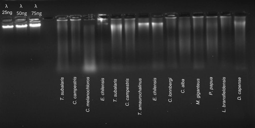
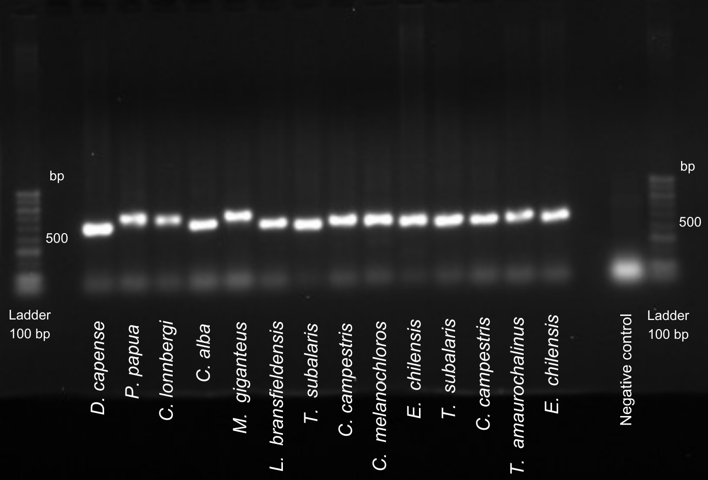
Cost analysis highlighted significant savings, with the protocol costing $0.05-$0.10 per sample compared to $2.50-$3.00 for commercial kits, such as the ReliaPrep™ gDNA Tissue Miniprep System. This difference represents a saving of up to 98%, making the reagent-based protocol a viable option for large-scale studies or in laboratories with budget constraints. For comparison purposes, DNA was also extracted using the ReliaPrep™ gDNA Tissue Miniprep System (Promega). Some degree of degradation was observed in both the samples isolated using the protocol developed in this study and those extracted using the commercial kit. However, the samples extracted with the commercial kit showed better integrity. In the samples extracted using the protocol developed, no significant differences were observed between the samples collected in 2024 and those collected between 1998-1999. Notably, the oldest sample, corresponding to Daption capense and stored for 26 years, showed integrity comparable to the most recent samples and to those extracted with the commercial kit (Figure 1). Despite the degree of degradation observed and the low amount of DNA obtained (Figure 1), these factors did not prevent efficient PCR amplification. The expected amplicon of around 559 bp was detected with good resolution in all the samples analyzed (Figure 2).
Discussion
DNA obtained from cell suspensions represents a practical and viable alternative for genomic studies, particularly when derived from samples originally prepared for cytogenetic analyses, as demonstrated by Amorim et al. (2007). Contrary to the findings of Nogueira and Freitas (2013), this study shows that the cell lysis technique is an effective method for extracting avian DNA from fixed materials, offering significant advantages over commercial kits due to its affordability. This methodology is especially beneficial for challenging scenarios, such as accessing samples from remote regions like Antarctica or rare specimens, including endangered species.
The use of fixed material samples provides both scientific and ethical benefits, allowing genetic studies to proceed without the need for new sample collections. This aligns with research ethics guidelines that emphasize reducing the number of animals used in scientific studies. Leveraging biological material stored for extended periods reduces the impact on populations of endangered species and adheres to the 3Rs principles (replacement, reduction, and refinement) (Hubrecht and Carter, 2019; Díaz et al., 2020). These principles play a crucial role in minimizing animal suffering and decreasing the reliance on new animal collections for research purposes.
However, extracting DNA from Farmer-fixed samples presents inherent challenges. Cells treated with hypotonic solutions and fixatives such as methanol and acetic acid become structurally fragile and susceptible to contamination, potentially compromising DNA quality and hindering analyses like PCR. Residues from heparin and the culture medium can persist despite thorough washing, interfering with PCR efficiency depending on their concentration (Schrader et al., 2012). Nevertheless, in this study, no significant interference from heparin or residual culture medium was observed, as corroborated by Beránek et al. (2022). These authors reported that heparin, when present in appropriate concentrations, interferes less with PCR reactions compared to other anticoagulants like EDTA and citrate. Floridia et al. (2023) further emphasized that variations in anticoagulants, including heparin, minimally affect gene expression in quantitative PCR, highlighting their applicability in diverse experimental contexts.
The low protein content observed in the extracted samples is attributed to the denaturing effects of the fixative and the action of Proteinase K, which degrades proteins during the extraction process. Prolonged storage can exacerbate genetic material degradation; however, no significant differences were noted between recently collected samples and those stored since 1998. Proper maintenance of the fixative is essential, as inadequate replacement can compromise cell preservation and further degrade DNA. These findings highlight the importance of strict storage and handling conditions to maintain the integrity of genetic material and minimize degradation impacts on subsequent analyses.
Conclusion
This study validates the viability of extracting DNA from Farmer-fixed avian cells for molecular research, demonstrating significant cost and ethical advantages. The proposed method effectively extracts DNA from Farmer-fixed avian cells, offering a cost-effective alternative to commercial kits. Its application aligns with ethical research principles, reducing the need for new sample collections and minimizing ecological impact. Despite degradation challenges, the protocol consistently yielded DNA suitable for PCR, emphasizing its potential for cytogenomic studies. Additionally, adherence to strict storage conditions can further enhance DNA integrity in future studies. By utilizing existing biological collections, this approach provides a sustainable framework for advancing genetic and evolutionary studies in avian species.
Acknowledgments
The authors would like to thank the Coordenação de Aperfeiçoamento de Pessoal de Nível Superior (CAPES), Conselho Nacional de Desenvolvimento Científico e Tecnológico (CNPq - grant 407285/2021-0), (A.D.V.G.), and FAPERGS for funding support.
Statement of ethics
The study protocol was reviewed and approved by the Ethics Committee on the Use of Animals, under approval numbers CEUA 019/2020 and CEUA 024/2023. Additional approvals were obtained from the Biodiversity Authorization and Information System (SISBIO), with authorization numbers 61047-3, 33860-2, and 81564-1. Some samples were collected with authorization from the Argentine Antarctic Institute.
Funding sources
This research was supported by FAPERGS through the Program for Supporting the Retention of Young PhDs in Brazil (EDITAL FAPERGS/CNPq 07/2022, awarded to L.O.M.) and by the National Council for Scientific and Technological Development (CNPq), Grant Number 407285/2021-0 (A.D.V.G.).
Author contributions
L.O.M., A.V.G., and R.J.G. contributed to the conception and design of the study. Methodology was carried out by L.O.M. and L.R.P. Formal analysis, data curation, and investigation were conducted by L.O.M. and F.P.T. Visualization was prepared by L.O.M. and A.S.K. The original draft was written by L.O.M. and H.S.S. Review and editing were performed by H.S.S., L.R.P., F.P.T., A.S.K., A.V.G., and R.J.G. Funding acquisition was handled by R.J.G. and A.V.G.
References
Amorim MR, Vargas FR, Llerena Junior JC, Pombo-de-Oliveira MS (2007). DNA extraction from fixed cytogenetics cell suspensions. Genet Mol Res 6(3):500–503.
Beránek M, Hegerová J, Michajlíková M, Dulíček P, Palicka V (2022). No inhibitory effect of heparinized blood on real-time PCR analysis of thrombophilic mutations. Folia Biol (Praha) 68:201–205. https://doi.org/10.14712/fb2022068050201.
Díaz C, Carmona E, Nuñez R, et al. (2020). Ethics in biological sample preservation: Applications and challenges in wildlife research. Biol Conserv 250:108–120.
Díaz L, Zambrano E, Flores ME, et al. (2020). Ethical considerations in animal research: The principle of 3R’s. Rev Invest Clin 73(4):199–209. https://doi.org/10.24875/RIC.20000380.
Floridia M, Pafundi S, Cecchi C, et al. (2023). Comparative effects of fixatives on RNA and DNA integrity: Implications for molecular diagnostics. Diagn Mol Pathol.
Floridia V, Sfulcini M, D’Alessandro E, et al. (2023). Effect of different anticoagulant agents on immune-related genes in leukocytes isolated from the whole-blood of Holstein cows. Genes (Basel) 14(2):406. https://doi.org/10.3390/genes14020406.
Garnero AV, Gunski RJ (2000). Comparative analysis of the karyotype of Nothura maculosa and Rynchotus rufescens (Aves: Tinamidae): A case of chromosomal polymorphism. Nucleus 43:64–70.
Grimm H, Biller-Andorno N, Buch T, et al. (2023). Advancing the 3Rs: innovation, implementation, ethics and society. Front Vet Sci 10:1185706. https://doi.org/10.3389/fvets.2023.1185706.
Hubrecht R, Carter E (2019). The 3Rs and the ethics of animal research. Anim Res Ethics 12(3):45–58.
Kalkan K, Kekecoglu M (2024). Ethical principles and rules in experimental animal studies: A comprehensive review. Düzce Tıp Fakültesi Dergisi 26. https://doi.org/10.18678/dtfd.1497730.
Kulkarni MD, Shringarpure R, Bhatt P, Prakash N, Prakash V (2015). A simple and inexpensive protocol for DNA isolation from avian blood. J Bombay Nat Hist Soc 112:3–7. https://doi.org/10.17087/jbnhs/2015/v112i1/92186.
Lott JA, Coleman WB, Tsongalis GJ, eds. (1997). Clinical Pathology of Pancreatic Disease and Molecular Diagnostics: For the Clinical Laboratorian. 1st ed. [s.l.]: [s.n.].
Miyaki CY (2001). Genética e evolução aplicada à conservação. In: Ornitologia e Conservação. Da Ciência às Estratégias. Editora Unisul, pp. 239–246.
Moorhead PS, Nowell PC, Mellman WJ, Battips DM, Hungerford DA (1960). Chromosome preparations of leukocytes cultured from human peripheral blood. Exp Cell Res 20:613–616. https://doi.org/10.1016/0014-4827(60)90138-5.
Nogueira DM, Freitas AAR (2013). Fixed cytogenetic cells suspension: An alternative for obtaining DNA of birds. Rev Bras Ornitol 21:120–123.
Pereira MA, Dias AR, Faraj SF, Cirqueira CS, Tomitao MT, Nahas SC, Ribeiro U Jr, de Mello ES (2015). Histopathology 66:388–397. https://doi.org/10.1111/his.12532.
Schrader C, Schielke A, Ellerbroek L, Johne R (2012). PCR inhibitors – occurrence, properties and removal. J Appl Microbiol 113(5):1014–1026.
Srinivasan M, Sedmak D, Jewell S (2002). Effect of fixatives and tissue processing on the content and integrity of nucleic acids. Am J Pathol 161(6):1961–1971. https://doi.org/10.1016/S0002-9440(10)64472-0.
Tan SC, Yiap BC (2009). DNA, RNA, and protein extraction: The past and the present. J Biomed Biotechnol 2009:1–10.
Supplement material
| Species | Extraction Protocol | A260/A280 | A260/A230 | Concentration (ng/ µl) |
|---|---|---|---|---|
| Daption capense | lysis | 1.6 | 1.8 | 236.5 |
| Pygoscelis papua | lysis | 1.5 | 2.1 | 466 |
| Catharacta lonnbergi | lysis | 1.6 | 1.8 | 2424 |
| Chionis alba | lysis | 1.6 | 1.9 | 713 |
| Macronectes giganteus | lysis | 1.7 | 2.0 | 1163 |
| Leucocarbo bransfieldensis | lysis | 1.7 | 2.0 | 1558 |
| Turdus subalaris | lysis | 1.7 | 1.6 | 1890 |
| Colaptes campestris | lysis | 1.5 | 1.5 | 2009 |
| Colaptes melanochloros | lysis | 1.6 | 1.5 | 368.5 |
| Elaenia chilensis | lysis | 1.7 | 1.8 | 1126 |
| Turdus subalaris | Commercial Kit | 1.6 | 1.7 | 417 |
| Colaptes campestris | Commercial Kit | 1.8 | 1.8 | 1138 |
| Turdus amaurochalinus | Commercial Kit | 2.1 | 3.0 | 42 |
| Elaenia chilensis | Commercial Kit | 1.8 | 1.9 | 161.5 |