Received: March 20, 2025; Accepted: November 10, 2025; Published: December 24, 2025
Lead and copper toxicity affecting chromosome structure, cell death, and micronucleus formation in Glycine max Cv-JS-355 root tip cells
Department of Biology, College of Science, King Khalid University, Abha 61413, Saudi Arabia
Email: sasdeky@kku.edu.sa
Abstract. The rapid rise of heavy metals and their extensive industrial use have raised concerns because these metals are released into the environment from both intentional and unintentional sources. When present in the environment in high concentrations, heavy metals may threaten the plant kingdom, particularly staple food crops. Nevertheless, little research has been done to identify the effects of heavy metals. The current study aims to assess the cytological alterations caused by lead (Pb) and copper (Cu) heavy metals on Glycine max Cv-JS-355. For two hours, Glycine max seeds were subjected to different Pb and Cu concentrations (CN, 25, 50, 75, 100, and 125 ppm). They were examined for their effects on chromosomal aberrations (CAs), micronucleus index (MNI), radicle length (RL), mitotic index (MI), cell death (CD), and seed germination (SG). The findings show a dose-dependent rise in MNI, CAs, CD and a substantial decrease in SG, RL, and MI. Furthermore, the percentage of abnormal mitotic cells, including cell nucleic leaking (CNL), Multi-pole division (MPD), Chromosomal bridge at telophase (CBT), chromosome retarded in anaphase (CRA), Dissociate chromosome in metaphase (DCM), increased in the Pb and Cu treated groups.
Keywords: Heavy metals (Pb and Cu), seed germination, radicle length, mitotic index, genotoxicity, cell death, Glycine max Cv-JS-355.
Contents
Glycine max seeds and chemicals procurement
Experiment site, seed germination, and radicle length analysis
Detection of mitotic index and chromosomal abnormality in root tips (RTs) of Glycine max
Detection of cell death in RTs of Glycine max
Detection of micronucleus index in RTs of Glycine max
Effect of heavy metals on SG of Glycine max
Effect of heavy metals on RL of Glycine max
Effect of heavy metals on MI of Glycine max
Effect of heavy metals on CAs of Glycine max
Effect of Pb and Cu on CD in RTCs of Glycine max
Effect of Pb and Cu on MNI in RTCs of Glycine max
Introduction
Heavy metals from mining, vehicle emissions, and agricultural effluents can pollute soil, affecting health of humans because of their noxious nature and higher bioaccumulation factor, potentially causing long-term ecosystem impacts (Dietrich et al. 2019; Neto et al. 2020). Heavy metal deficiency leads to environmental and toxicological issues due to increased contamination from industrialization and pesticide use. Contamination from agricultural waste, home sewage, and industrial discharges enters water bodies, reducing water quality and increasing metal availability in food chain (Hassan et al. 2020). Rapid industrialization and urbanization worldwide lead to the toxicity of dangerous metals in soil, including lead, copper, nickel, aluminum, and cadmium (Zhang et al. 2024), often resulting from irrigation from wastewater (Iqbal et al. 2016).
Increased levels of heavy metals in the soil have several detrimental impacts on plants, including decreased growth, inhibited root development, degradation of chlorophyll, altered biochemical activity, and reproductive diseases linked to oxidative damage (Colzi et al. 2015; Abdull et al. 2022). Furthermore, chromosomal irregularities, point mutations, and ploidy are among the genetic alterations that can result from the buildup of hazardous substances (Silveira et al. 2017). Programmed cell death (PCD), which is triggered by controlled intracellular signals, and nonprogrammed cell death as accidental cell death, which includes necrosis, are two types of cell death. Heavy metals are an example of an abiotic elemenst that can impact cells and cause necrosis or PCD (Brighigna 2006; van Doorn et al. 2011; Petrov et al. 2015).
Metals may have long-lasting negative effects on the ecosystem since they are non-biodegradable and can remain in soil for longer intervals of time. Former studies have shown that microorganisms present in soil, like plants growing in filthy locations, are vulnerable to high dosages of heavy metals, which can lead to malfunction, denaturation of protein, and compromised integrity of cell membranes (Hosseini et al. 2022). Accordingly, metals that are found in cationic forms interact with sulfhydryl radical (-SH) that are found in protein structures of enzymes, altering their characteristics and having detrimental effects on metabolism of plants (Nowicka 2022; Siddiqui 2025a). Metals play key functions in the metabolism of plants, and their characteristics are critical to the tridimensional maintenance of biomolecules and cell metabolism. Nevertheless, certain metals are required in trace amounts, others have no biological significance and may even cause metabolic harm (Neto et al. 2020).
In most countries around the world, Soybeans (Glycine max L.) are widely farmed for industrial, animal, and human purposes because of their higher protein and oil content (Siddiqui 2025b). Soybeans are frequently known as miracle crops since they are the world’s main protein and vegetable oil source. Considering the growing global population and the need for increased crop productivity, this study aims to evaluate the effects of Pb and Cu on Glycine max L, seed germination, radicle length, mitotic index, cell death, micronucleus index, and chromosomal abnormalities.
Methodology
Glycine max seeds and chemicals procurement
Certified soybean seeds of Glycine max L Merr. Cv-JS-355 were purchased from CSIR (Council of Scientific and Industrial Research), Bhopal, India. Sigma-Aldrich Company was a supplier of heavy metals CuSO4 and PbSO4.
Experiment site, seed germination, and radicle length analysis
From October to December, the tests were carried out at King Khalid University’s Botany Department, Al-Farra campus, Abha, Saudi Arabia. Six treatments of Pb and Cu were prepared: CN, 25, 50, 75, 100, and 125 ppm. The completely randomized methods comprise an experimental setup. In Petri dishes covered with a double layer of Whitman No. 2 paper, fifty Glycine max Cv-JS-355 seeds were planted. Ten milliliters of a solution with different Pb and Cu concentrations of CN, 25, 50, 75, 100, and 125 ppm were placed in each plate. Distilled water was taken as control (CN). The plates were maintained, with few adjustments, in a B.O.D. chamber (Cienlab®) at 18 ºC ± 2 ºC having a photo period of 12 hours, as per recommendations given by Siddiqui (2023). Seed germination was measured after 24, 48, and 72 h. Radicle length was measured after 24 h for three days with measuring scale.
Detection of mitotic index and chromosomal abnormality in root tips (RTs) of Glycine max
For cytogenetic assays, all plates were made utilizing the smashing technique, incorporating a slight modification as described by Siddiqui (2023). Root tips Glycine max were harvested after 72 hours of each treatment and subsequently fixed in a Carnoy’s solution (3 parts ethanol : 1 part glacial acetic acid) and preserved at a temperature of 18 °C. Three samples, each containing 150 cells, were assessed, resulting in a total of 450 cells per replicate. The behavior of chromosomes were analyzed by quantifying the cell cycle stages. MI was calculated based on number of cells undergoing mitosis relative to total number of observed cells. Chromosomal irregularities were evaluated using specific criteria: cell nucleic leaking, multi-pole division, chromosomal bridge in telophase, chromosome retarded in anaphase, dissociate chromosome in metaphase with their frequencies determined by the total number of abnormalities and the overall cell count.
Detection of cell death in RTs of Glycine max
To assess cell death in RTs, uptake of non-permeable trypan blue dye was utilized. Trypan blue can only penetrate the membrane of a dead cell. Using the procedure described by Duan et al. (2010), RTs (0.1 cm) were immersed in trypan blue (0.4%, w/v) for 15 minutes at room temperature. After that, they were twice rinsed with 2.5 g/mL chloral hydrate solution for ten minutes. Following sample preparation, pictures were captured, and analysis was done using an optical microscope (Olympus CX23, Japan).
Detection of micronucleus index in RTs of Glycine max
Each slide’s 100 cells were scored to calculate the MN for assessment. A light binocular microscope (Olympus) with a magnification of 100x was used to investigate micronucleated cells. For scoring MNI, the technique stated by Tolbert et al. (1992) was utilized.
Statistical analysis
A one-way ANOVA test was used to examine the significance of differences between variables using the GPIS 1.13 program (GRAPHPAD, California, USA). All results were presented as mean ± standard error.
Results
Effect of heavy metals on SG of Glycine max
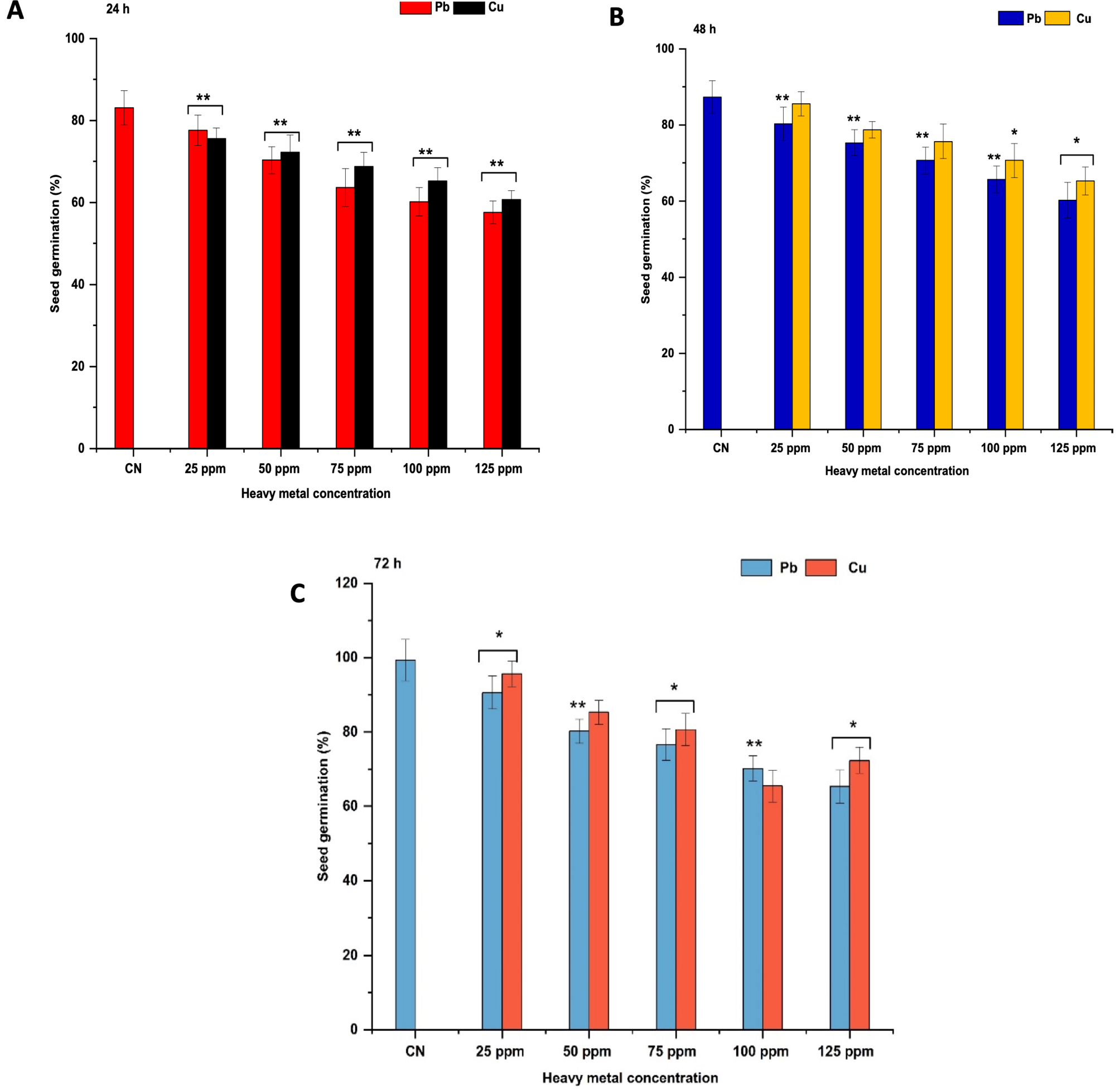
In untreated seeds, after 2 h, germination percentage of seeds were 83.12%, 87.3%, and 99.3% at 24, 48, and 72 h respectively (Figure 1. A-C). Pb and Cu treatments of 25 to 125 ppm for 2 h caused a very significant SG decline (p<0.01) at 24 h relative to control. This pattern remained the same for SG at 48 and 72 h, where highest SG was recorded at 25 ppm at 24 h (Pb: 77.66 %, Cu: 75.65 %), 48 h (Pb: 80.33 %, Cu: 85.54 %), and 72 h (Pb: 90.65 %, Cu: 95.67 %), on treating with Pb and Cu for 2 h while lowest SG was found at 125 ppm at 24 h (Pb: 57.63%, Cu: 60.76%), at 48 h (Pb: 60.25%, Cu: 65.27%), and at 72 h (Pb: 65.35 %, Cu: 72.35 %) relative to control.
Effect of heavy metals on RL of Glycine max
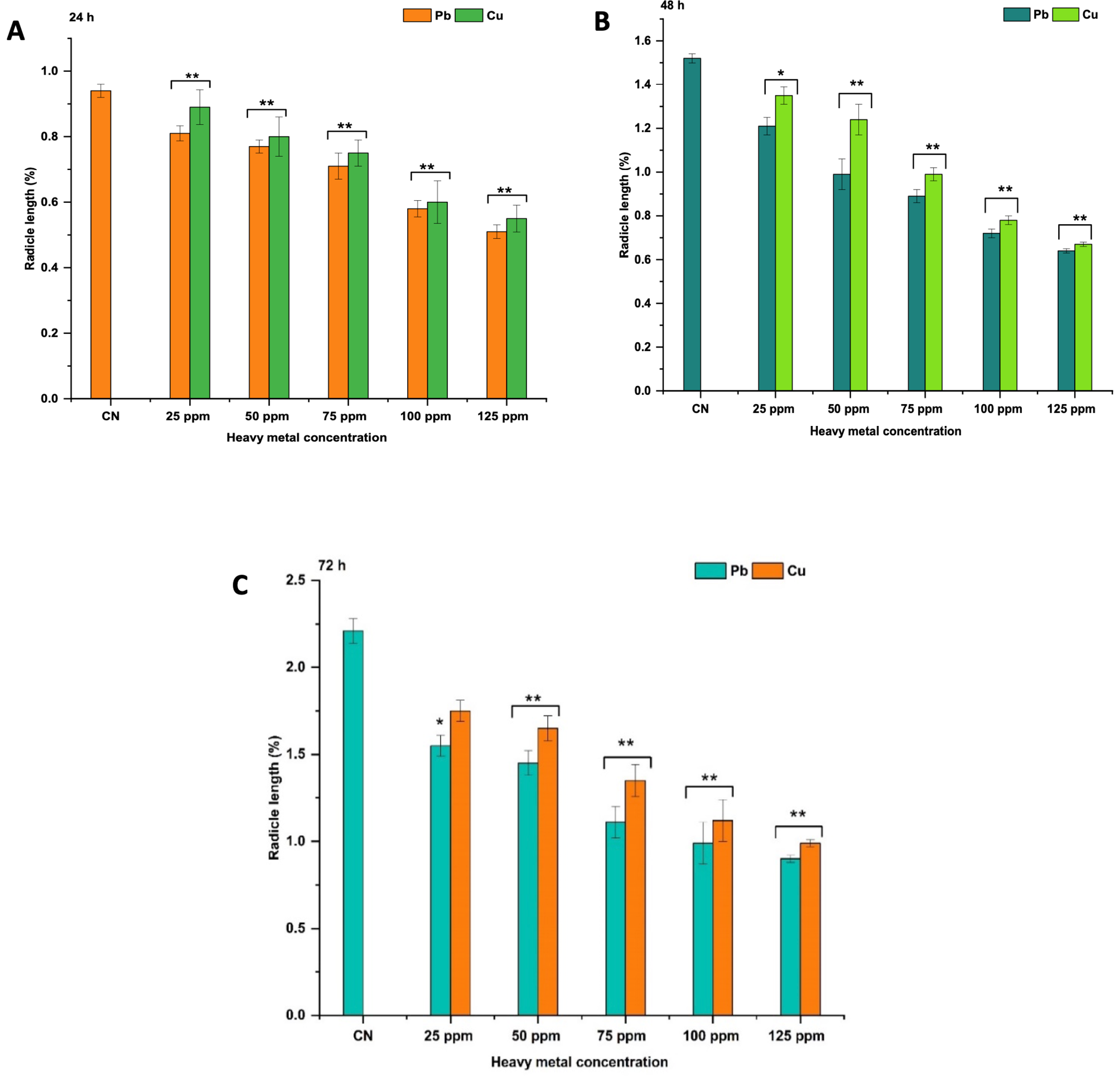
Pb and Cu effect on untreated seeds show that RL rose with time on treating with double-distilled water (DDW) for 2 h: 0.94±0.020 at 24 h, 1.52±0.021 at 48 h, and 2.21±0.071 at 72 h (Figure 2. A-C). Pb and Cu treatments of 25 ppm to 125 ppm for 2 h caused significant RL decline (p<0.05 at 25 ppm and p<0.01 at 50 to 125 ppm) relative to control. This pattern remained the same for RL at 48 h and 72 h, where highest RL was recorded at 25 ppm at 24 h (Pb: 0.81±0.023, Cu: 0.89± 0.053), at 48 h (Pb: 1.21±0.04, Cu: 1.35±0.04), and at 72 h (Pb: 1.55±0.06, Cu: 1.75±0.061) on treating with Pb and Cu for 2 h while lowest RL was found at 125 ppm at 24 h (Pb: 0.51±0.021, Cu: 0.55±0.041), at 48 h (Pb: 0.64±0.01, Cu: 0.67±0.01), and at 72 h (Pb: 0.90±0.02, Cu 0.99±0.02) relative to control.
Effect of heavy metals on MI of Glycine max
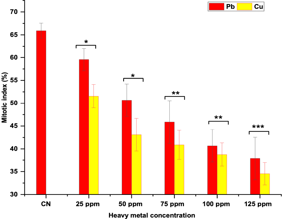
Figure 3 illustrates the impact of Pb and Cu on MI of root tip cells (RTCs) of Glycine max. In control, treatment with DDW for 2 h, exhibited MI values of about 65.90%. Seeds exposed to 25 ppm to 50 ppm of Pb and Cu for 2 h showed a significant reduction (p<0.05) in MI relative to control. A dose of 75 to 100 ppm of Pb and Cu for 2 h revealed a very significant reduction (p <0.01), while 125 ppm of Pb and Cu caused a highly significant reduction (p<0.001) in MI. Maximal MI was found at 25 ppm (Pb: 59.56%, Cu: 51.55%) and minimal MI was found at 125 ppm (Pb: 37.88%, Cu: 34.53%) relative to control.
Effect of heavy metals on CAs of Glycine max
No aberrant metaphase-anaphase plates were found in RTs of Glycine max in control (Table 1, Figure 4). Different types of CAs, such as cell nucleic leaking (CNL), multi-pole division (MPD), chromosomal bridge in telophase (CBT), chromosome retarded in anaphase (CRA), and dissociate chromosome in metaphase (DCM) were found in metaphase-anaphase plates. In seeds exposed to Pb and Cu for 2 h, there is a surge in ratio of aberrant metaphase-anaphase plates with a surge in Pb and Cu concentration. Cytological investigations disclose that level of CA steadily escalated with a surge in concentrations of Pb and Cu treatment for 2 hours. Investigations of varied stages of mitotic division reveal that all stages of the division were altered.
| Conc. (ppm) | CNL | MPD | CBT | CRA | DCM | ||||||||||||||
|---|---|---|---|---|---|---|---|---|---|---|---|---|---|---|---|---|---|---|---|
| CN | 0.00±0.00 | 0.00±0.00 | 0.00±0.00 | 0.00±0.00 | 0.00±0.00 | ||||||||||||||
| Pb | |||||||||||||||||||
| 25 ppm | 0.80±0.02* | 0.42±0.01* | 0.42±0.01* | 0.21±0.02 | 0.80±0.04* | ||||||||||||||
| 50 ppm | 1.20±0.04** | 0.80±0.24** | 0.31±0.24** | 0.80±0.24* | 0.80±0.02** | ||||||||||||||
| 75 ppm | 1.20±0.04* | 0.40±0.02* | 0.40±0.02* | 1.00±0.10* | 0.40±0.02** | ||||||||||||||
| 100 ppm | 1.60±0.37* | 0.80±0.10* | 0.20±0.01* | 0.40±0.01** | 1.22±0.40** | ||||||||||||||
| 125 ppm | 2.25±0.54** | 2.10±0.44*** | 0.40±0.01** | 0.80±0.04 *** | 1.23±0.21** | ||||||||||||||
| Cu | |||||||||||||||||||
| 25 ppm | 0.80±0.02* | 0.20±0.01 | 0.30±0.01* | 0.20±0.01* | 0.40± 0.06* | ||||||||||||||
| 50 ppm | 1.20±0.03* | 0.20±0.42 | 0.40±0.03* | 0.40±0.02* | 0.80±0.02* | ||||||||||||||
| 75 ppm | 1.20±0.03* | 0.80±0.21* | 0.20±0.05* | 0.20±0.01** | 1.20±0.24** | ||||||||||||||
| 100 ppm | 0.40±0.04** | 0.80±0.24** | 0.30±0.21** | 0.80±0.22** | 1.20±0.24** | ||||||||||||||
| 125 ppm | 1.20±0.15*** | 1.40±0.12*** | 1.40±0.15** | 0.40± 0.21*** | 2.12± 0.63*** | ||||||||||||||
| *P<0.05; **P<0.01; ***P<0.001 compared to control group. Data are mean of three replicates ± SE, CN = control group, where CNL (Cell nucleic leaking); MPD (Multi-pole division); CBT (Chromosomal bridge in telophase), CRA (chromosome retarded in anaphase), DCM (Dissociate chromosome in metaphase). | |||||||||||||||||||
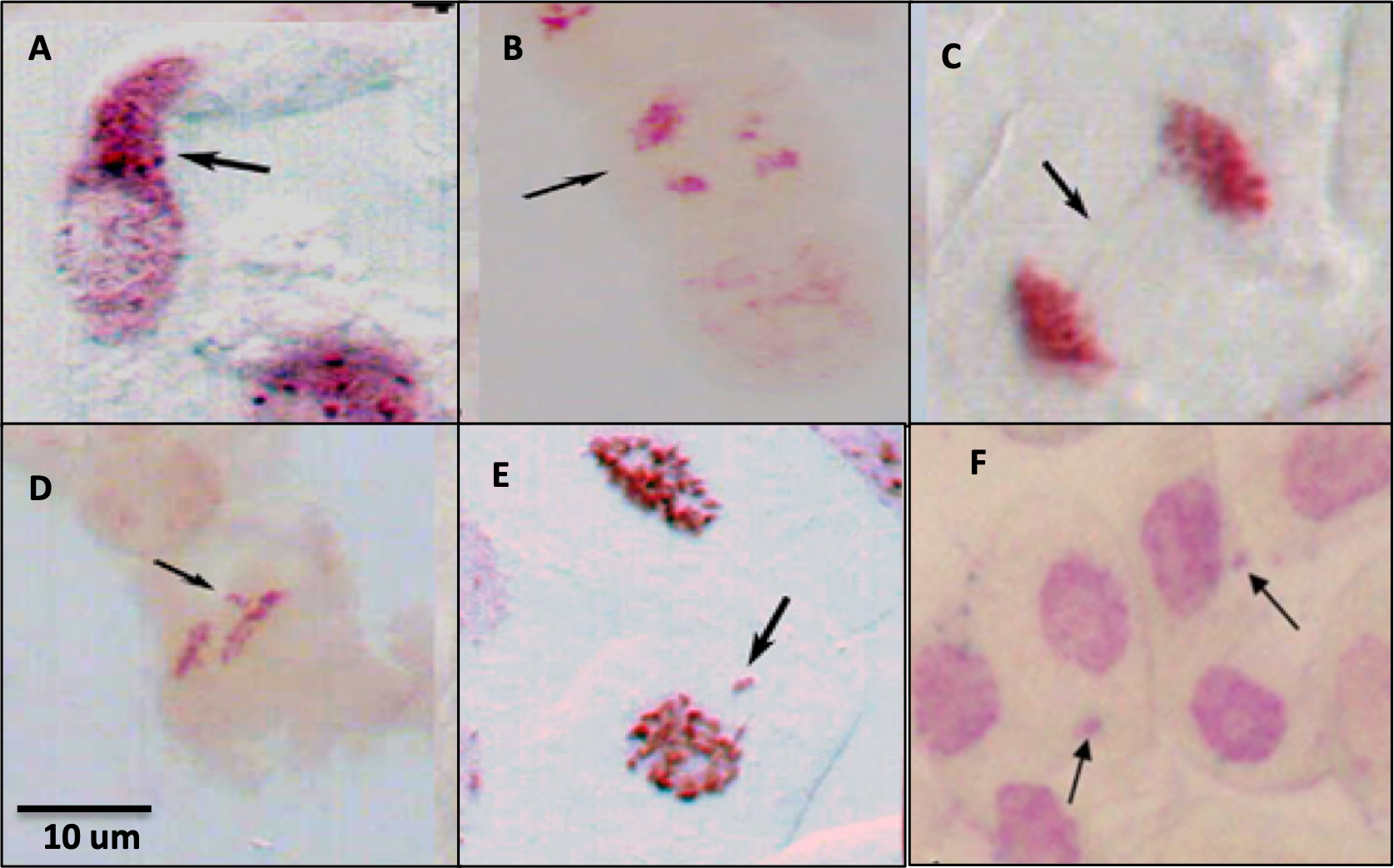
Percentage formation of CNL, MPD, CBT, CRA, and DCM were very significant (p<0.01) and maximal at 125 ppm such as CNL (2.25%), MPD (2.1%), CBT (0.40%), CRA (0.80%) and DCM (1.23%) after Pb treatment. Minimal percentage of chromosomal anomalies were at 25 ppm CNL (0.8%), CRA (0.21%), and MPD (0.40%), DCM (0.40%) at 75 ppm, CBT at 100 ppm (0.2%), at 2 h of Pb exposed seeds relative to control. Enhanced occurrence of CAs for Pb exposure were:
CNL > MPD >DCM >CRA> CBT
Similarly percentage formation of CNL, MPD, CBT, CRA, and DCM were very significant (p<0.01) and maximal at 125 ppm such as CNL (1.20%), MPD (1.40%), DCM (2.12%), and CBT (1.40%), and CRA (0.80% ) at 100 ppm at 2 h of Cu treated seeds. Minimal percentage of chromosomal anomalies were reported at 100 ppm CNL (0.4%), MPD at 25 and 50 ppm (0.20%), BRT at 75 ppm (0.2%), CRA at 25 and 75 ppm (0.2%) and DCM at 25 ppm (0.40%) at 2 h of Cu exposed seeds relative to control. Enhanced occurrence of CAs for Cu exposure were:
DCM >MPD = CBT > CNL > CRA
Effect of Pb and Cu on CD in RTCs of Glycine max
Figure 5 illustrates the impact of Pb and Cu on CD in Glycine max RTCs. In control, treatment with DDW for 2 h, exhibited CD of 0.11%. Seeds exposed to 25 ppm of Pb for 2 h exhibited a nonsignificant rise in CD which was 1.40% in comparison to control. Seeds exposed to 50 ppm for 2 h with Pb exhibited a significant rise (p<0.05) in CD which was 2.80% relative to control. At 75 ppm to 100 ppm, a very significant rise (p<0.01) in CD was revealed which was 3.75% at 75 ppm and 4.90% at 100 ppm relative to control. At 125 ppm, a highly significant rise (p<0.001) in CD was revealed which was 5.85 % relative to control. Minimal CD was found at 25 ppm (1.40%), and maximal CD was found at 125 ppm (5.85%). Rise in CD was dose dependent.
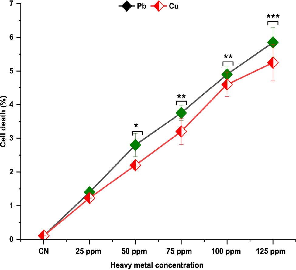
Seeds exposed to 25 ppm of Cu for 2 h exhibited a nonsignificant rise in CD which was 1.23% relative to control. Seeds exposed to 50 ppm for 2 h with Cu exhibited a significant rise (p<0.05) in CD which was 2.2% relative to control. At 75 ppm to 100 ppm a very significant rise (p<0.01) in CD was revealed which was 3.2% at 75 ppm and 4.6% at 100 ppm as compared to control. At 125 ppm a highly significant rise (p<0.001) in CD was revealed which was 5.25% in comparison to control. Minimal CD was found at 25 ppm (1.23%), and maximal CD was found at 125 ppm (5.25%). The rise in CD was dose dependent.
Effect of Pb and Cu on MNI in RTCs of Glycine max
Figure 6 illustrates the impact of Pb and Cu on MNI of Glycine max in RTCs. In control, MNI was 0.15% of RTCs treated with DDW for 2 h. A significant rise (p<0.05) in MNI was found in RTCs treated for 2 h with Pb at 25 ppm which was 0.80% and very significant rise (p<0.01) in MNI was found at 50, 75 and 100 ppm which were 1.80 %, 2.80 %, and 3.92% respectively, and highly significant rise (p<0.001) in MNI was found at 125 ppm which was 5.23%, in comparison to control. Minimal MNI was found at 25 ppm (0.80%), and maximal MNI was found at 125 ppm (5.23%). Rise in MNI was dose dependent.
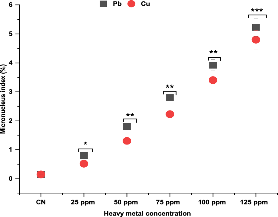
A significant rise (p<0.05) in MNI was found in RTCs treated for 2 h with Cu at 25 ppm which was 0.52% and a very significant rise (p<0.01) in MNI was found at 50, 75 and 100 ppm which were 1.30%, 2.23%, and 3.40% respectively and highly significant rise (p<0.001) in MNI was found at 125 ppm which was 4.80%, in comparison to control. Minimal MNI was found at 25 ppm (0.52%), and maximal MNI was found at 125 ppm (4.80%). Rise in MNI was dose dependent.
Discussion
This study noticed delayed SG, RL, MI, increased CD, MNI, and CA in Glycine max root tip cells. According to previous research, Pb and Cu reduces germination (Siddiqui et al. 2009; Siddiqui 2013; Sarac et al. 2019; Nouri et al 2019) and causes toxicity and mutagenicity in various plant species (da Cunha Neto 2020; Siddiqui and Sulaiman 22021; da Cunha Neto et al. 2023). When seeds are sown in high Cd environments, their ability to survive is hampered, preventing the development of the embryonic axis and radicle. This impacts activity of α and β amylases (Karmous et al. 2015). Numerous factors are known to affect seed germination, including light, moisture content, oxygen concentration, and incubation temperature (Siddiqui 2024 a and b; Anwar et al. 2025). It has been demonstrated that heavy metals prevent seeds from germinating at higher concentrations (Siddiqui 2012; 2015; Akbaş 2024). A decrease in RL is thought to be caused by heavy metal-induced root cell death, specifically because of inadequate energy synthesis in root cells (Siddiqui 2018; Siddiqui et al. 2021; Tasar et al. 2022; Qin et al. 2024). In Brassica napus seedlings, heavy metals caused cell death and formation of callose in addition to lowering the micronutrient concentrations (Pramanik et al. 2018).
MI is a crucial factor in determining the genotoxic potential of heavy metals. In Glycine max, cytotoxicity elucidates delay in growth, whereas genotoxicity explicates chromosomal abnormalities. Pesticides, heavy metals and other chemical pollutants exposure were linked to the decline in MI in Pisum sativum (Siddiqui et al. 2007; Siddiqui et al. 2020 a and b, 2022 a, b and c). Delays in cell cycle or chromatin disorder brought on by metal-DNA interaction causes a decrease in MI (Siddiqui et al. 2012; Ditika and Anila 2013). During cell division, heavy metal affects mitosis and causes spindle-related chromosomal aberrations (Siddiqui 2012; 2015). Numerous findings have demonstrated that differences in the mitotic cycle’s duration may be the cause of a decline in cell activity. A rise in the S phase’s duration has been attributed by some researchers to mitotic inhibition (Siddiqui et al. 2012; Periakaruppan et al. 2023). As per Das et al. (2023), heavy metals can reach DNA by nuclear pores or when nuclear membrane splits into cells undergoing mitotic divide.
Our results show that CD in the RTs of Glycine max exposed to heavy metals at all doses from 25, 50, 75, 100, and 125 ppm increases in a dose-dependent manner. For programmed cell death, plants use vacuolar content and vacuoles in two ways: destructively and non-destructively (Yang et al. 2023). Direct and immediate cell death results from the release of vacuolar hydrolytic enzymes into the cytoplasm following the rupture of the vacuolar membrane (Brighigna et al. 2006). As organelles that generate energy, mitochondria help cells divide, operate, and elongate by metabolizing energy (Fenech et al. 2011; Siddiqui 2024a).
Presence of micronuclei in the root tip cells of Glycine max further confirmed the genotoxicity caused by heavy metals. After two hours of exposure to heavy metals concentration being increased from 25 to 125 ppm, they significantly increased the number of MNI. The precise mechanism by which heavy metals induce the formation of micronuclei is still unknown. Conversely, micronuclei form during mitosis because of lagging chromosomes or acentric fragments. (Fenech et al. 2011; Yan 2011; Sabeen et al. 2020). Heavy metal-induced damage to parental cells that are either unrepaired or improperly healed and may have a mutagenic effect (Shobha et al 2020). Similar genotoxic effects of heavy metals on Triticum sativum, Glycine max, Vicia faba, and Allium cepa have been reported by other studies (Bapi et al. 2018; Abdelkader et al. 2022).
The formation of chromosomal aberrations are a useful way to analyze genotoxicity. To assess the extent of chromosomal damage, this study looked at changes in chromosomal behavior brought on by heavy metals. Although the control plant lacked CAs, treated plants showed several abnormalities, such as cell nucleic leaking, multi-pole division, chromosomal bridge at telophase, chromosome retarded in anaphase, dissociate chromosome in metaphase even at the lowest dosage. The frequency of these abnormalities suggests that they have aneugenic, tubergenic, and clastogenic effects (Siddiqui 2019; Liu et al. 2021; Abdelsalam et al. 2022). They are brought on by chromosomal breakage (fragments, micronuclei) and spindle apparatus disorders, anomalous metaphase, telophase, anaphase, and bridges (Zhang et al. 2024; Leng et al. 2025)
Conclusion
The dosage of exposure to heavy metals was directly correlated with their cytotoxicity. It is suggested that ambient heavy metals are readily absorbed and finally move up the food chain and higher concentrations of these heavy metals may harm plant by inducing chromosomal aberrations, cell death, alterations in mitotic index, micronucleus index radicle length, and seed germination. Additionally, they impede plant growth by causing oxidative stress, heavy metals have the potential to harm the ecosystem by causing DNA damage. These results are important for the proper disposal of heavy metals and the levels at which they become dangerous.
Funding
The authors extend their appreciation to the Deanship of Research and Graduate Studies at King Khalid University for funding this work through the Large Group Project under grant number (RGP2/89/46)
References
Abdelkader M, Geioushy RA, Fouad OA, Khaled AG. 2022. Investigation of the activities of photosynthetic pigments, antioxidant enzymes and inducing genotoxicity of cucumber seedling exposed to copper oxides nanoparticles stress. Sci. Hortic. 305:111364.
Abdelsalam NR, Abdel-Megeed A, Ghareeb RY, Ali HM, Salem MZ, Akrami M, Al-Hayalif, MF, Desoky ESM. 2022. Genotoxicity assessment of amino zinc nanoparticles in wheat (Triticum aestivum L.) as cytogenetical perspective. Saudi J. Biol. Sci. 29: 2306–2313.
Abdull SR, Rashid SH, Ghafoor BS, Khdhir BS. 2022. Effect of Ag Nanoparticles on morphological and physio-biochemical traits of the medicinal plant Stevia rebaudiana. Caryologia. 75(2): 15–22.
Akbaş. 2024. effects of different concentrations OF Cu+, Mn+, and Ni+ ions on Glycine max germination. Bilim Armonisi. 7(2): 26–36.
Anwar A, Akhtar J, Aleem S, Aleem M, Razzaq MK, Alamri S, Raza Q, Sharif I, Iftikhar A, Naseer S, Ahmed Z. 2025. Genome-wide identification of MGT gene family in soybean (Glycine max) and their expression analyses under magnesium stress conditions. BMC Plant Biology. 25(1): 83.
Bapi G, Kumar DA, Debadrito D, Vishambhar KD, Ankita P. 2018. Assessment of nanoparticles (copper, cadmium sulphide, copper oxide and zinc oxide) mediated toxicity in a plant system (Indigofera tinctoria L.; Fabaceae). Res. J. Chem. Environ. 22: 34–48.
Brighigna L, Papini A, Milocani E, Vesprini JL. 2006. Programmed cell death in the nucellus of Tillandsia (Bromeliaceae). Caryologia. 59(4): 334-9.
Colzi I, Pignattelli S, Giorni E, Papini A, Gonnelli C. 2015. Linking root traits to copper exclusion mechanisms in Silene paradoxa L.(Caryophyllaceae). Plant and Soil. 390(1): 1–5.
da Cunha Neto AR, Carvalho M, Morais GM, Guaraldo MM, Dos Santos HO, Pereira WV, Barbosa S. 2023. Changes in chromosome complement and germination of lettuce (Lactuca sativa L.) exposed to heavy metal stress. Water, Air, & Soil Pollution. 234(4): 243.
da Cunha Neto, AR, Ambrósio, ADS, Wolowski, M, Westin TB, Govêa, KP, Carvalho, M, Barbosa, S. 2020. Negative effects on photosynthesis and chloroplast pigments exposed to lead and aluminum: A meta-analysis. CERNE. 26(2): 232–37. https://doi.org/10.1590/01047 760202026022711
Das D, Bisht K, Chauhan A, Gautam S, Jaiswal JP, Salvi P, Lohani P. 2023. Morpho-physiological and biochemical responses in wheat foliar sprayed with zinc-chitosan-salicylic acid nanoparticles during drought stress. Plant Nano Biol. 4: 100034.
Dietrich M, Wolfe A, Burke M, Krekeler MPS. 2019. The first pollution investigation of road sediment in Gary, Indiana: anthropogenic metals and possible health implications for a socioeconomically disadvantaged area. Environ. Int. 128: 175–192. https://doi.org/10.1016/J.ENVINT.2019.04.042
Ditika K, Anila M. 2013. Assessment of cytotoxic and genotoxic potency of Cr (VI)-doped river water of Nen-Shkodra lowland, Albania, on Allium cepa L. J Environ. Res. Dev. 7(4): 1322–1332
Duan Y, Zhang W, Li B, Wang Y, Li K, Sodmergen, HC, Li X. 2010. An endoplasmic reticulum response pathway mediates programmed cell death of root tip induced by water stress in Arabidopsis. New Phytol. 186: 681–695. https://doi.org/10.1111/j.1469-8137.2010.03207.x
Fenech M, Krisch-Volders M, Natarajan AT, Surralles J, Crott JW, Parry J, Noppa H, Eastmond DA, Tucker JD, Thomas P. 2011. Molecular mechanism of micronucleus, nucleoplasmic bridge and nuclear bud formation in mammalian and human cells. Mutagenesis. 26: 125–132.
Hassan MM, Haleem N, Baig MA, JamalY. 2020. Phytoaccumulation of heavy metals from municipal solid waste leachate using different grasses under hydroponic condition. Sci. Rep. 10(1): 1–8. https://doi.org/10.1038/s41598-020-72800-2
Hosseini, S. M., Kalatejari, S., Kafi, M., & Motesharezadeh, B. (2023). Assessment of the absorption ability of nitrate and lead by japanese raisin under salt stress conditions. Caryologia 75(4). https://doi.org/10.36253/caryologia-1827
Iqbal HH, Taseer R, Anwar S, Qadir A, Shahid N. 2016. Human health risk assessment: heavy metal contamination of vegetables in Bahawalpur, Pakistan Hafiza. Bull. Environ. Stud. 1(1): 10–17
Karmous I, Bellani LM, Chaoui A, El Ferjani E, Muccifora S. 2015. Effects of copper on reserve mobilization in embryo of Phaseolus vulgaris L. Environ. Sci. Pollut. Res. 22(13): 10159–10165. https://doi.org/10.1007/S11356-015-4208-1/FIGURES/7
Leng Y, Niu ZB, Liu SH, Qiao FJ, Liu GF, Cheng B, Li SW. 2025. Characterisation of cytochrome c oxidase-coding genes from mung bean and their response to cadmium stress based on genome-wide identification and transcriptome analysis. Mol. Biol. Rep. 52(1): 17.
Liu C, Yu Y, Liu H, Xin H. 2021. Effect of different copper oxide particles on cell division and related genes of soybean roots. Plant Physiol. Biochem. 163: 205–14.
Neto AR, da Ambrósio C, Wolowski ADS, Westin M, Govêa TB, Carvalho KP, Barbosa MS. 2020. Negative effects on photosynthesis and chloroplast pigments exposed to lead and aluminum: a meta-analysis. CERNE 26(2): 232–237. https://doi.org/10.1590/01047760202026022711
Nouri M, El Rasafi T, Haddioui A. 2019. Responses of two barley subspecies to induced heavy metal stress: seeds germination, seedlings growth and cytotoxicity assay. Agriculture 65(3) 107–18.
Nowicka B. 2022. Heavy metal–induced stress in eukaryotic algae—Mechanisms of heavy metal toxicity and tolerance with particular emphasis on oxidative stress in exposed cells and the role of antioxidant response. Environ. Sci. Pollut. Res. 29(12): 16860–16911. https://doi.org/10.1007/s11356-021-18419-w
Periakaruppan R, Vanathi P, Priyanka G, Vidhya D. 2023. Toxicity in plants by metal oxide nanoparticles. In Nanometal Oxides in Horticulture and Agronomy; Academic Press: Cambridge, MA, USA. 241–273.
Petrov V, Hille J, Mueller-Roeber B, Gechev TS. 2015. ROS-mediated abiotic stress-induced programmed cell death in plants. Front. Plant Sci. 6: 69.
Pramanik A, Datta AK, Gupta S, Ghosh B. 2018. Copper oxide nanoparticles induced fertile desynaptic mutant line in Coriandrum sativum L. (Apiaceae). Cytologia. 83: 103–107.
Qin C, Lian H, Alqahtani FM, Ahanger MA. 2024. Chromium-mediated damaging effects on growth, nitrogen metabolism and chlorophyll synthesis in tomato can be alleviated by foliar application of melatonin and jasmonic acid priming. Sci. Hortic. 323: 112494.
Sabeen M, Mahmood Q, Bhatti ZA, Irshad M, Bilal M, Hayat MT, Irshad U, Akbar TA, Arslan M, Shahid N. 2020. Allium cepa assay-based comparative study of selected vegetables and the chromosomal aberrations due to heavy metal accumulation. Saudi J. Biol. Sci. 27(5): 1368–4.
Sarac I, Bonciu E, Butnariu M, Petrescu I, Madosa E. 2019. Evaluation of the cytotoxic and genotoxic potential of some heavy metals by use of allium test. Caryologia. 72(2): 37–43. https://doi.org/10.13128/cayologia-256.
Shobha G, Shashidhara KS, Naik C. 2020. Cuprous oxide nanoparticles induced antioxidant response and genotoxicity in Lycopersicum esculentum. Bio Nanosci. 10: 1128–1137
Siddiqui S, 2023. Phenthoate toxicity evaluation in root meristem of Pisum sativum L. Caryologia. 76(1): 57–66.
Siddiqui S, Al Amri SAM, Al Ghamdy HA, Alqahtani WSS, Alquyr SM, Yassin HM. 2021. Impact of Bisphenol A on seed germination, radicle length and cytogenetic alterations in Pisum sativum L. Caryologia. 74(2): 103–109.
Siddiqui S, Al-Rumman S. 2020a. Clethodim induced pollen sterility and meiotic abnormalities in vegetable crop Pisum sativum L. Caryologia. 73: 37–44.
Siddiqui S, Al-Rumman S. 2020b. Cytological changes induced by clethodim in Pisum sativum plant. Bangladesh J. Bot. 49(2): 367–374.
Siddiqui S, Al-Rumman S. 2022a. Methomyl, imbraclaobrid and clethodim induced cytomixis and syncytes behaviors in PMCs of Pisum sativum L: Causes and outcomes. Saudi J Biol Sci. 29(9): 103390. https://doi.org/10.1016/j.sjbs.2022.103390.
Siddiqui S, Al-Rumman S. 2022b. Exposure of Pisum sativum L. seeds to methomyl and imidacloprid cause genotoxic effects in pollen-mother cells. Biology. 11: 1549. https://doi.org/10.3390/ biology11111549
Siddiqui S, Al-Rumman S. 2022c. Methomyl has clastogenic and aneugenic effects and alters the mitotic kinetics in Pisum sativum L. Caryologia. 75(3): 91–99.
Siddiqui S, Meghvansi MK, Hasan Z. 2007. Cytogenetic changes induced by sodium azide (NaN3) on Trigonella foenum-graecum L. seeds. S. Afr. J. Bot. 73(4): 632–5.
Siddiqui S, Meghvansi MK, Khan SS. 2012. Glyphosate, alachor and maleic hydrazide have genotoxic effect on Trigonella foenum-graecum L. Bull. Environ. Contam. Toxicol. 88(5): 659-65.
Siddiqui S, Meghvansi MK, Wani MA, Jabee F. 2009. Evaluating cadmium toxicity in the root meristem of Pisum sativum L. Acta Physiol. Plant. 31: 531–6.
Siddiqui S. 2012. Lead-induced genotoxicity in Vigna mungo var. HD-94. J. Saudi Soc. Agric. Sci. 11(2): 107–12.
Siddiqui S. 2013. Exposure of Cu and Mn to Cicer arietinum L. Var. BGD-72 seeds induces morphological and biochemical changes in the plant. South Asian J. Exp. Biol. 3(1): 31–36.
Siddiqui S. 2015. DNA damage in Cicer plant grown on soil polluted with heavy metals. J. King Saud Univ. Sci. 27(3): 217–23.
Siddiqui S. 2018. Cytotoxicity induced by aluminum sulfate in cells of root meristem of Pisum sativum cv. arikil. Bangl. J. Bot. 1: 47:219.
Siddiqui S. 2024a. DNA Damage, cell death, and alteration of cell proliferation insights caused by copper oxide nanoparticles using a plant-based model. Biology. 13(10): 805.
Siddiqui S. 2024b. Effects of cypermethrin on morphological, physiological and biochemical attributes of Cicer arietinum (Fabales: Fabaceae). Front. Sustain. Food Syst. 8: 1446308.
Siddiqui S. 2025a. Global patterns and drivers of species and genera richness of Fabaceae. Front. Plant Sci. 16: 1581814.
Siddiqui S. 2025b. Unlocking the environmental potential of biochar: production, applications, and limitations. Frontiers in Sustainable Food Systems. 9: 1569941
Silveira GL, Lima MGF, Reis GB, Palmieri MJ, Andrade-Vieria LF. 2017. Toxic effects of environmental pollutants: a comparative investigation using Allium cepa L. and Lactuca sativa L. Chemosphere. 178: 359–367. https://doi.org/10.1016/J.CHEMOSPHERE.2017.03.048
Tasar N. 2022. Mitotic effects of copper oxide nanoparticle on root development and root tip cells of Phaseolus vulgaris L. seeds. Microsc. Res. Tech. 85: 3895–3907.
Tolbert PE, Shy CM, Allen JW. 1992. Micronuclei and other nuclear anomalies in buccal smears: Methods development. Mutat. Res. 271: 69–77.
van Doorn WG, Beers EP, Dangl JL, Franklin-Tong VE, Gallois P, Hara-Nishimura I, Jones AM, Kawai-Yamada M, Lam E, Mundy J, Mur LA. 2011.Morphological classification of plant cell deaths. Cell Death Differ. 18(8): 1241–6.
Zhang Y, Li T, Fu Q, Hou R, Li M, Liu D, Shi G, Yang X, Xue P. 2024. Drip irrigation reduces the toxicity of heavy metals to soybean: By moving heavy metals out of the root zone and improving physiological metabolism. Agric. Water Manag. 292: 108670