https://doi.org/10.36253/caryologia-3431
Received: March 26, 2025; Accepted: July 17, 2025; Published: December 20, 2025
Karyotype analysis and chromosome evolution in Menyanthaceae using FISH
Department of Applied Plant Science, College of Agriculture and Life Sciences, Kangwon National University, Chuncheon 24341, Republic of Korea
*Corresponding author. E-mail: laurus@kangwon.ac.kr
Abstract. The Menyanthaceae, an aquatic plant family, is distinguished by extensive polyploidy and heterostyly. This study marks the first cytogenetic characterization of four Menyanthaceae species from Korea – Menyanthes trifoliata, Nymphoides peltata, N. indica, and N. coreana – employing fluorescence in situ hybridization (FISH) with 45S and 5S rDNA probes. All four species exhibit exclusively metacentric chromosomes, with M. trifoliata and N. peltata being hexaploid (2n = 54), N. coreana tetraploid (2n = 36), and N. indica diploid (2n = 18). FISH mapping revealed between one to four 45S rDNA loci and one to three 5S rDNA loci per species, showing that rDNA site number does not correlate directly with ploidy level. The karyotypic data suggest a conserved base chromosome number (x = 9) and largely symmetrical karyotypes across these species. Notably, M. trifoliata presents fewer rDNA loci than expected for a hexaploid, indicating genomic rearrangements and rDNA locus loss through diploidization. These observations highlight an evolutionarily stable genome structure in M. trifoliata, despite its polyploid nature. This study elucidates the chromosomal organization and evolutionary dynamics of the Menyanthaceae, emphasizing the role of polyploidy and rDNA evolution in genome structuring. The findings enhance our understanding of plant cytogenetics in aquatic ecosystems and serve as a foundation for further comparative genomic and evolutionary studies in Menyanthaceae.
Keywords: Menyanthaceae, polyploidy, karyotype analysis, Fluorescence in situ hybridization, rDNA loci.
Contents
Root sample preparation for chromosome
Fluorescence in situ hybridization (FISH)
Genetic evolutionary dynamics in Menyanthaceae
Genomic status of Menyanthes trifoliata
Discrepancy between FISH signals and ploidy level
Introduction
The Menyanthaceae family comprises perennial, floating-leaved aquatic plants prevalent across pantropical regions, including tropical America, Asia, and Australia. These plants are noted for heterostyly, a self-incompatible reproductive system that encourages outcrossing (Barrett 1992; Barrett and Shore 2008). The family predominantly exhibits distyly, though variants such as homostyly or other mating system alterations occur (Ornduff 1970, 1987, 1992). Reproduction is facilitated through both sexual (seed-based) and asexual (clonal propagation via root-derived turions) modes, promoting ecological resilience and adaptability in wetland habitats (Tippery et al. 2008, 2009). In the Korean Peninsula, Menyanthaceae is represented by Menyanthes trifoliata L. and Nymphoides peltata (S. G. Gmel.). Kuntze, Nymphoides indica (L.) Kuntze, and Nymphoides coreana (H. Lév.) H. Hara, all crucial for biodiversity and ecological stability in aquatic environments (Ornduff 1970; Tippery et al. 2008, 2009; Watanabe 2022).
Understanding chromosomal organization in Menyanthaceae is crucial for unraveling their genetic and evolutionary dynamics, especially given their adaptation to aquatic environments. Cytogenetic studies have revealed substantial chromosomal variation within this family, including phenomena such as polyploidy, descending dysploidy, and chromosomal races, which contribute to speciation and environmental adaptation (Gillett 1968; Watanabe 2022; Leitch and Leitch 2013). In the genus Nymphoides (x = 9), detailed examinations reveal chromosome counts predominantly in diploid (2n = 18), tetraploid (2n = 36), and hexaploid (2n = 54) states, indicating dynamic karyotypic evolution (Ornduff 1970). Similarly, M. trifoliata shows a hexaploid chromosome count (2n = 54), contrasting with Nephrophyllidium crista-galli, which has been documented with 2n = 108, highlighting extensive polyploidization within the Menyanthaceae (Gillett 1968; Tippery et al. 2008).
Fluorescence in situ hybridization (FISH) is a robust cytogenetic technique that facilitates the direct visualization of specific DNA sequences on chromosomes (Abbo et al. 1994). This method uses fluorescently labeled probes to map vital genomic elements, such as 45S and 5S rDNA loci, which act as molecular markers for assessing chromosomal polymorphisms and genetic diversity (Stebbins 1971; Stace 2000; Ilnicki 2014). Recent studies have underscored the role of rDNA loci in chromosomal evolution, particularly among plant species undergoing polyploidization and structural rearrangements (Weiss-Schneeweiss et al. 2013; Watanabe 2022).
This study aims to investigate the chromosomal architecture of M. trifoliata using FISH to map repetitive DNA sequences, including 45S and 5S rDNA loci. By analyzing the karyotype, we hope to elucidate the organization, composition, and evolutionary dynamics of the Menyanthaceae genome. Comparative analyses with related taxa will provide deeper insights into chromosomal evolution and adaptation strategies in wetland environments, contributing to broader knowledge in plant cytogenetics, genome stability, and conservation biology.
Material and methods
Root sample preparation for chromosome
Plant materials were collected from natural populations (Table 1). Roots were pre-treated with 2 mM 8-hydroxyquinoline solution for 4 hours at 12°C. They were subsequently fixed in Carnoy’s solution (3:1 ethanol: acetic acid) for 24 hours and stored in 70% ethanol at 4°C until use.
| Taxa | Collection data |
|---|---|
| Menyanthes trifoliata L. | Korea, Gangwon-do, Goseong-gun, April 13, 2021, H. R. Kim and K. Heo s.n. (KWNU) |
| Korea, Gangwon-do, Taebaek-si, May 25, 2021, H. R. Kim and K. Heo s.n. (KWNU) | |
| Nymphoides indica (L.) Kuntze | Korea, Jeju-do, Jocheon-eup, August 8, 2023, K. Heo s.n. (KWNU) |
| Korea, Jeju-do, Jocheon-eup, August 12, 2023, K. Heo s.n. (KWNU) | |
| Nymphoides coreana (H.Lév.) H.Hara | Korea, Jeju-do, Seogwipo-si, July 30, 2023, H. R. Kim and J. S. Yang s.n (KWNU) |
| Nymphoides peltata (S.G.Gmel.) Kuntze | Korea, Gyeonggi-do, Yangpyeong-gun, September 11, 2022, H. R. Kim and K. Heo s.n. (KWNU) |
Chromosome spread preparation
Somatic chromosome spreads were prepared using a modified version of the technique described by Kirov et al. (2014). After thorough washing with distilled water, the meristematic regions of the fixed root tips were excised and digested in an enzyme mix (2% cellulase, 1% pectolyase in 1× Citrate buffer) for 52 minutes at 37°C. The enzyme mix was removed, and 80 μL of Carnoy’s solution was added; roots were suspended by vortexing, then centrifuged, and the pellet was resuspended in a 9:1 acetic acid–ethanol solution. Finally, the root suspension was dropped onto slides in a humid chamber to spread the chromosomes, and then the chromosomes were air-dried.
Fluorescence in situ hybridization (FISH)
The 45S rDNA was labeled with digoxigenin-11-dUTP (Roche, Germany) via nick translation and detected with anti-digoxigenin FITC. The 5S rDNA was labeled with biotin-16-dUTP and detected with streptavidin–avidin Cy3 (Table 2). Labeled DNA fragments ranging from 100 to 500 bp were used as probes. The hybridization mixture for FISH contained 50% formamide, 10% dextran sulfate, 2×SSC, and 500 ng/μL of each probe DNA, adjusted with distilled water to a total volume of 50 μL per slide. The mixture was denatured at 90°C for 10 minutes and immediately cooled on ice for 10 minutes. After applying the probe mixture, chromosome slides were denatured at 80°C for 3 minutes on a hotplate. The slides were then incubated in a humid chamber at 37°C for 18 hours to facilitate hybridization. Subsequently, the slides were treated with 2x SSC for 5 minutes at RT and 1x detection buffer for 10 minutes at RT. Biotin‐labelled 5S rDNA and digoxigenin labelled 45S rDNA were detected using Cy3-conjugated streptavidin and anti-digoxigenin-fluorescein isothiocyanate (FITC) at 37°C for 1 hour. Excess reagents were removed by washing three times in 1x detection buffer for 5 minutes each. The slides were dehydrated in a series of ethanol (70%, 90%, and 100%) for 3 minutes at RT, and air-dried. Then, the slides were counterstained with DAPI in VECTASHIELD. Chromosome spreads were examined using a phase-contrast fluorescence microscope (Axio Imager M2, Carl Zeiss, Germany). Chromosome length measurements and image acquisition were performed with ZEN software (Carl Zeiss).
| Gene | Type | Sequence |
|---|---|---|
| 5s rDNA | Forward | 5’-CGGTGCATTAATGCTGGTAT-3’ |
| Reverse | 5’-CCATCAGAACTCCGCAGTTA-3’ | |
| 45s rDNA | Forward | 5’-CGAAACCTGCAAGAGCA-3’ |
| Reverse | 5’-GTCTGATCTGGGGTCGCAA-3’ |
Results
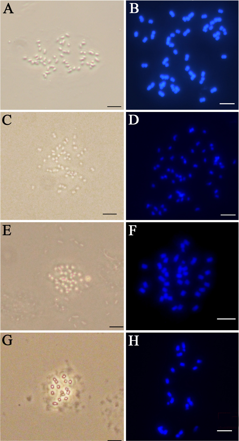
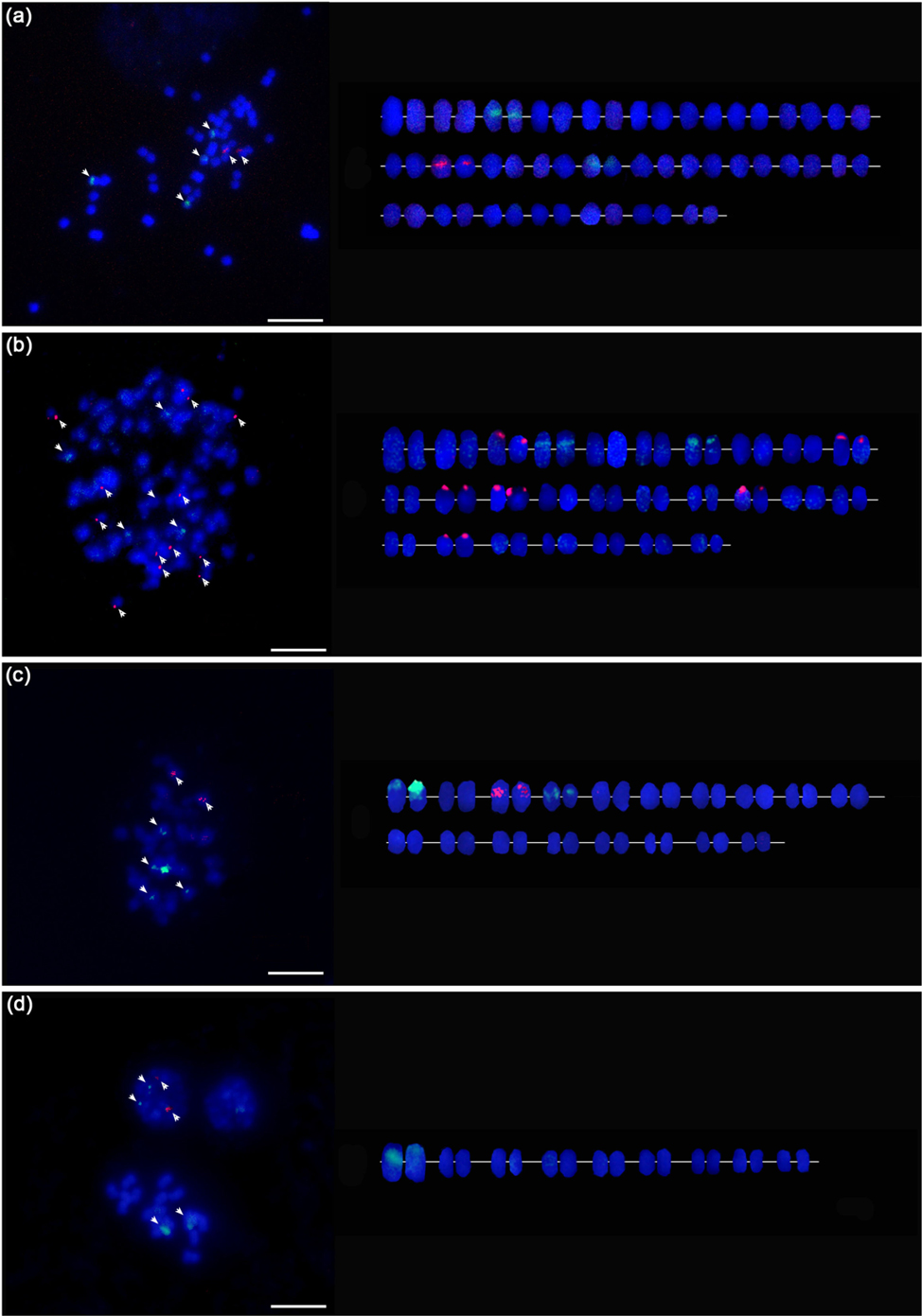
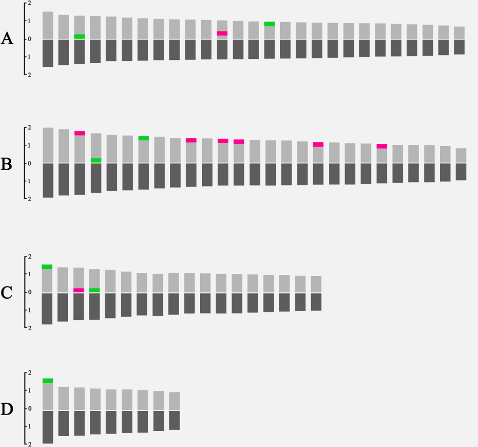
This study provides the first cytogenetic characterization of four Menyanthaceae species using double-FISH. All species were found to possess exclusively metacentric chromosomes (Figs 1, 3). Chromosome counts confirm that M. trifoliata and N. peltata are hexaploid (2n = 54, Fig. 2A, B), N. coreana is tetraploid (2n = 36, Fig. 2C), and N. indica is diploid (2n = 18, Fig. 2D). Chromosome lengths varied from approximately 1.0 μm to 3.7 μm across these four species (Table 3; Fig. 3). FISH mapping identified between one and four 45S rDNA loci and one to three 5S rDNA loci per species, indicative of some variation in rDNA copy number among the genomes (Fig. 2.).
| Ch. No | M. trifoliata | N. peltata | N. coreana | N. indica | |||||||||||||||
|---|---|---|---|---|---|---|---|---|---|---|---|---|---|---|---|---|---|---|---|
| CL (μm) | AR (μm) | Type | CL (μm) | AR (μm) | Type | CL (μm) | AR (μm) | Type | CL (μm) | AR (μm) | Type | ||||||||
| (mean±SD) | (mean±SD) | (mean±SD) | (mean±SD) | (mean±SD) | (mean±SD) | (mean±SD) | (mean±SD) | ||||||||||||
| 1 | 3.01±0.37 | 1.14±0.05 | m | 3.88±0.45 | 1.04±0.10 | m | 3.26±0.28 | 1.04±0.05 | m* | 3.71±0.30 | 1.14±0.06 | m° | |||||||
| 2 | 2.82±0.35 | 1.12±0.05 | m | 3.68±0.33 | 1.12±0.07 | m | 2.87±0.33 | 1.12±0.12 | m | 2.80±0.33 | 1.12±0.06 | m | |||||||
| 3 | 2.57±0.26 | 1.05±0.06 | m* | 3.06±0.38 | 1.11±0.05 | m° | 2.70±0.33 | 1.11±0.10 | m° | 2.75±0.22 | 1.05±0.04 | m | |||||||
| 4 | 2.42±0.22 | 1.08±0.08 | m | 3.31±0.31 | 1.03±0.13 | m* | 2.68±0.34 | 1.03±0.07 | m* | 2.61±0.25 | 1.08±0.07 | m | |||||||
| 5 | 2.34±0.22 | 1.09±0.04 | m | 3.20±0.36 | 1.07±0.06 | m | 2.56±0.38 | 1.07±0.05 | m | 2.52±0.14 | 1.09±0.04 | m | |||||||
| 6 | 2.27±0.18 | 1.08±0.06 | m | 3.17±0.57 | 1.07±0.10 | m | 2.52±0.39 | 1.07±0.13 | m | 2.47±0.28 | 1.08±0.05 | m | |||||||
| 7 | 2.21±0.17 | 1.09±0.09 | m | 3.13±0.28 | 1.01±0.05 | m* | 2.52±0.17 | 1.08±0.06 | m | 2.42±0.50 | 1.09±0.10 | m | |||||||
| 8 | 2.17±0.16 | 1.09±0.05 | m | 2.95±0.50 | 1.04±0.08 | m | 2.46±0.22 | 1.04±0.10 | m | 2.27±0.22 | 1.09±0.08 | m | |||||||
| 9 | 2.14±0.15 | 1.10±0.09 | m | 2.08±0.22 | 1.11±0.04 | m | 2.41±0.28 | 1.12±0.05 | m | 2.14±0.17 | 1.10±0.08 | m | |||||||
| 10 | 2.10±0.16 | 1.11±0.07 | m | 2.79±0.37 | 1.06±0.03 | m° | 2.37±0.60 | 1.06±0.08 | m | ||||||||||
| 11 | 2.04±0.16 | 1.08±0.07 | m | 2.75±0.19 | 1.09±0.04 | m | 2.35±0.54 | 1.09±0.04 | m | ||||||||||
| 12 | 1.99±0.14 | 1.08±0.04 | m° | 2.68±0.25 | 1.01±0.02 | m° | 2.31±0.31 | 1.01±0.03 | m | ||||||||||
| 13 | 1.96±0.14 | 1.07±0.09 | m | 2.64±0.23 | 1.06±0.05 | m° | 2.25±0.19 | 1.06±0.03 | m | ||||||||||
| 14 | 1.93±0.13 | 1.09±0.10 | m | 2.61±0.17 | 1.01±0.05 | m | 2.23±0.25 | 1.01±0.05 | m | ||||||||||
| 15 | 1.91±0.13 | 1.06±0.04 | m* | 2.58±0.15 | 1.05±0.06 | m | 2.22±0.25 | 1.07±0.02 | m | ||||||||||
| 16 | 1.88±0.14 | 1.09±0.12 | m | 2.56±0.60 | 1.14±0.06 | m | 2.22±0.19 | 1.14±0.05 | m | ||||||||||
| 17 | 1.85±0.13 | 1.10±0.03 | m | 2.50±0.33 | 1.06±0.05 | m | 2.21±0.36 | 1.06±0.07 | m | ||||||||||
| 18 | 1.83±0.12 | 1.09±0.02 | m | 2.45±0.16 | 1.00±0.03 | m° | 2.02±0.19 | 1.00±0.04 | m | ||||||||||
| 19 | 1.79±0.12 | 1.11±0.02 | m | 2.38±0.15 | 1.14±0.07 | m | |||||||||||||
| 20 | 1.76±0.12 | 1.12±0.06 | m | 2.38±0.12 | 1.04±0.04 | m | |||||||||||||
| 21 | 1.72±0.11 | 1.10±0.05 | m | 2.30±0.19 | 1.08±0.05 | m | |||||||||||||
| 22 | 1.67±0.11 | 1.06±0.03 | m | 2.17±0.27 | 1.11±0.07 | m° | |||||||||||||
| 23 | 1.63±0.12 | 1.10±0.09 | m | 2.11±0.22 | 1.07±0.10 | m | |||||||||||||
| 24 | 1.59±0.10 | 1.12±0.06 | m | 2.09±0.23 | 1.04±0.05 | m | |||||||||||||
| 25 | 1.52±0.10 | 1.11±0.10 | m | 2.06±0.14 | 1.12±0.03 | m | |||||||||||||
| 26 | 1.46±0.09 | 1.09±0.05 | m | 1.97±0.20 | 1.08±0.10 | m | |||||||||||||
| 27 | 1.37±0.07 | 1.10±0.04 | m | 1.86±0.24 | 1.10±0.13 | m | |||||||||||||
| CL: Chromosome length, AR: Arm ratio, SD: Standard deviation, m: Metacentric chromosome, *: 45s rDNA, °:5s rDNA. | |||||||||||||||||||
The precise chromosomal positions of the rDNA signals were determined for each species. In M. trifoliata, green fluorescence signals corresponding to 45S rDNA were detected on chromosomes 3 and 15, while red fluorescence signals for 5S rDNA were observed on chromosome 12. In N. peltata, 45S rDNA signals were detected on chromosomes 4 and 7, and 5S rDNA signals on chromosomes 3, 10, 12, 13, 23, and 27. N. coreana exhibited 45S rDNA signals on chromosome 3 and 5S rDNA signals on chromosomes 1 and 4. N. indica displayed a 45S rDNA signal on chromosome 1 and a single 5S rDNA signal, which exact position could not be determined (Table 3; Fig. 3).
Discussion
Genetic evolutionary dynamics in Menyanthaceae
The evolutionary trajectory of Menyanthaceae is characterized by extensive chromosomal variation and a prevalence of polyploidization, a key driver of speciation and morphological diversification. For example, the retention or breakdown of heterostyly can influence gene flow and mating patterns within Menyanthaceae populations (Haddadchi 2013, 2015; Barrett and Shore 2008). Studies on Villarsia (Menyanthaceae) reveal distinct stigma morphology differences between distylous and non-heterostylous species, supporting the role of reproductive adaptations in lineage diversification (Dulberger and Ornduff 2000).
Cytogenetic studies have consistently revealed a base chromosome number of x = 9 across the family (Ornduff 1970), with species exhibiting diploid (2n = 18), tetraploid (2n = 36), and hexaploid (2n = 54) karyotypes (Cook 1996). The genus Nymphoides exemplifies this pattern, wherein polyploidization appears to have enhanced ecological adaptability and geographic expansion (Soltis and Soltis 2016). Similar polyploidy-associated genomic modifications have been observed in other aquatic plant groups (Martel et al. 2004; Watanabe 2022).
Polyploidy is a recurrent feature in angiosperm evolution, yet its interaction with descending dysploidy remains an active area of research (Kadereit 2007). At the molecular level, FISH has provided critical insights into chromosomal evolution, especially concerning the organization of ribosomal DNA (rDNA) loci. Studies have revealed substantial variation in both the number and chromosomal positioning of rDNA loci across Nymphoides species, suggesting that post-polyploidization genomic reorganization is common (Rosato et al. 2015; Silvestri et al. 2015). Additionally, evidence of descending dysploidy – where chromosome number is reduced following polyploidization – indicates that Menyanthaceae species undergo structural karyotypic modifications to stabilize their genomes (Semple and Watanabe 2023). This ongoing genomic reorganization underscores the dynamic evolutionary landscape of the family, where a whole-genome duplication (WGD) is often followed by selective gene loss and structural rearrangements.
Genomic status of Menyanthes trifoliata
Despite cytogenetic confirmation that Menyanthes trifoliata is a hexaploid species with 2n = 54 chromosomes (Peruzzi and Cesca 2004), its genetic behavior raises fundamental questions about whether it functions as a true hexaploid or has undergone extensive diploidization. Unlike certain polyploid Nymphoides species displaying cytotype diversity, M. trifoliata has remained cytogenetically uniform across its widespread circumboreal distribution. This stability suggests it may represent an ancient hexaploid lineage that has functionally reverted to a diploid-like state through genomic restructuring (Raabová et al. 2010).
Several lines of evidence support the hypothesis that M. trifoliata has undergone diploidization. First, cytogenetic analyses indicate predominantly bivalent chromosome pairing during meiosis, characteristic typically associated with diploid-like inheritance (Mlinarec et al. 2012). Second, its rDNA organization deviates from what would be expected in a simple hexaploid genome, with fewer detectable rDNA loci than a direct tripling of the diploid number (Rosato et al. 2015). These patterns suggest that genomic streamlining has eliminated redundant rDNA arrays, favoring a more functionally efficient karyotype. Third, its consistent chromosome number across various geographic populations, with no evidence of aneuploidy or unstable cytotypes, further supports the notion of an evolutionarily stable genome structure (Soltis and Soltis 2016). Similar cases of diploidization have been observed in other polyploid plant taxa, including Nicotiana allopolyploids, where rDNA homogenization has played a role in genomic stabilization (Kovarik et al. 2008).
The precise origins of M. trifoliata remain unresolved. It may have arisen from autopolyploidy due to successive WGD events, or from allopolyploidy, in which hybridization between distinct ancestral genomes contributed to its karyotype (Watanabe 2022). Comparative genomic studies of M. trifoliata and closely related Nymphoides species could offer deeper insight into whether its hexaploid genome originated from hybridization or from independent lineage expansion (Watanabe 2022). Advances in high-throughput sequencing technologies will be key in resolving this issue by enabling a comprehensive analysis of genome duplication patterns and homeologous gene retention. Importantly, these genomic and cytogenetic observations can be interpreted within the framework of existing molecular phylogenies. Phylogenetic analyses based on chloroplast DNA and nuclear ITS regions have consistently placed Menyanthes as sister to a clade of polyploid Nymphoides species (Tippery et al. 2008; Watanabe 2022). Our FISH-based findings support this phylogenetic position by providing cytogenetic evidence that complements molecular data. Specifically, M. trifoliata exhibits diploid-like chromosomal behavior during meiosis and possesses fewer rDNA loci than would be expected under a strict hexaploid model. These features are consistent with a scenario in which a whole-genome duplication event occurred before the divergence of Menyanthes, followed by substantial genomic reorganization and diploidization. Furthermore, the extensive chromosomal variation observed among Nymphoides species aligns with their high level of molecular divergence, suggesting that polyploidy and subsequent chromosomal restructuring have played a major role in driving diversification within the genus. Thus, the cytogenetic patterns revealed in this study provide a structural and evolutionary context that complements and reinforces existing phylogenetic hypotheses for Menyanthaceae.
Discrepancy between FISH signals and ploidy level
An intriguing anomaly in Menyanthaceae cytogenetics is the absence of a direct correlation between the number of rDNA signals (as detected by FISH) and the ploidy level. Theoretically, a polyploid lineage derived from a diploid ancestor should display a proportional increase in rDNA loci. Yet, M. trifoliata exhibits fewer rDNA signals than expected for a strict hexaploid model (Fultz and Pikaard 2023). This discordance suggests that polyploid genomes undergo significant restructuring following duplication, leading to selective retention, loss, or relocation of rDNA loci. Notably, similar observations have been made in certain polyploid Solanaceae, where fewer rDNA loci are present than expected for their ploidy level.
One plausible explanation for this discrepancy is the selective loss of redundant rDNA loci. Polyploidization often results in an initial surplus of rDNA copies, but genome evolution may favor the retention of only the most functionally necessary loci, leading to the eventual elimination of extraneous rDNA sites (Mlinarec et al. 2012). A targeted analysis using quantitative PCR or whole-genome sequencing could help determine whether the observed reduction in rDNA FISH signals corresponds to actual sequence loss. Another contributing factor could be rDNA transposition and homogenization. In some polyploids, rDNA loci are not static; instead, they may undergo concerted evolution, where a subset of rDNA sites expands while others diminish or relocate to different chromosomes (Rosato et al. 2015). This pattern is also observed in maize, where rDNA transposition significantly influences chromosomal architecture (Li and Arumuganathan 2001). Such processes may explain why M. trifoliata exhibits a lower-than-expected number of 45S and 5S rDNA loci despite its hexaploid genome structure. Employing FISH with additional chromosomal markers, such as probes for transposable elements, could reveal whether rDNA sites have been repositioned within the genome. Epigenetic modifications, particularly nucleolar dominance, further complicate the relationship between rDNA loci and ploidy. In allopolyploids and some autopolyploids, nucleolar dominance can result in the silencing of rDNA loci from one parental genome, leading to a functional reduction in active rDNA sites despite their genomic presence (Fultz and Pikaard 2023). If M. trifoliata exhibits such a mechanism, certain rDNA loci may not be transcriptionally active, making them undetectable by FISH. RNA-seq analyses of rRNA transcription levels could elucidate whether epigenetic silencing contributes to the observed reduction in rDNA signals. The inconsistency between FISH signal number and ploidy level underscores the complexity of genome evolution in polyploids. Rather than a straightforward duplication of all genetic elements, polyploid genomes undergo intricate modifications, including chromosomal rearrangements, rDNA loss, and epigenetic regulation. Future research integrating molecular cytogenetics, high-resolution sequencing, and transcriptomic analyses will be essential to fully characterize the evolutionary dynamics of M. trifoliata and other polyploid Menyanthaceae taxa.
Acknowledgments
This study was supported by the Korea Research Foundation (KRF) with a grant under project number RS-2021-NR065781 (2021R1I1A2043432).
References
Abbo, S., Miller, T. E., Reader, S. M., Dunford, R. P. and King, I. P. 1994. Detection of ribosomal DNA sites in lentil and chickpea by fluorescent in situ hybridization. – Genome 37: 713–716.
Barrett, S. C. H. 1992. Heterostylous genetic polymorphisms: Model systems for evolutionary analysis. – Am. Nat. 139: 421–435.
Barrett, S. C. H. and Shore, J. S. 2008. New insights on heterostyly: Comparative biology, ecology and genetics. – Philos. Trans. R. Soc. B 363: 491–508.
Cook, C. D. K. 1996. Aquatic Plant Book. SPB Academic Publishing, Amsterdam.
Dulberger, R. and Ornduff, R. 2000. Stigma morphology in distylous and non-heterostylous species of Villarsia (Menyanthaceae). – Plant Syst. Evol. 225: 171–184.
Fultz, D. and Pikaard, C. S. 2023. Reaching for the off switch in nucleolar dominance. – Plant J. 115: 642–652.
Gillett, J. M. 1968. The systematics of the Asiatic and American populations of Fauria crista-galli (Menyanthaceae). – Can. J. Bot. 46: 92–96.
Haddadchi, A. 2013. Stylar polymorphism, reciprocity and incompatibility systems in Nymphoides montana (Menyanthaceae) endemic to southeastern Australia. – Plant Syst. Evol. 299: 389–401.
Haddadchi, A. and Fatemi, M. 2015. Self-compatibility and floral traits adapted for self-pollination allow homostylous Nymphoides geminata (Menyanthaceae) to persist in marginal habitats. – Plant Syst. Evol. 301: 239–250.
Ilnicki, T. 2014. Cytogenetic analysis of selected species using fluorescence in situ hybridization (FISH). – Caryologia 67: 192–196.
Kadereit, J. W. 2007. Menyanthaceae. In: Kadereit, J.W. and Jeffrey, C. (eds.), The Families and Genera of Vascular Plants, vol. 8. Springer, Berlin, pp. 225–242.
Kirov, I., Divashuk, M., Van Laere, K., Soloviev, A., and Khrustaleva, L. 2014. An easy “SteamDrop” method for high quality plant chromosome preparation. – Molecular Cytogenetics. 7: 1–10.
Kovarik, A., Dadejova, M., Lim, Y. K., Chase, M. W., Clarkson, J. J., Knapp, S. and Leitch, A. R. 2008. Evolution of rDNA in Nicotiana allopolyploids: a potential link between rDNA homogenization and epigenetics. – Ann. Bot. 101: 815–823.
Leitch, I. J. and Leitch, A. R. 2013. Genome size diversity and evolution in land plants. – New Phytol. 200: 3–18.
Li, L. J. and Arumuganathan, K. 2001. Physical mapping of sorted chromosomes 45S and 5S rDNA on maize metaphase by FISH. – Hereditas 134: 141–145.
Martel, E., Poncet, V., Lamy, F., Siljak-Yakovlev, S., Lejeune, B. and Sarr, A. 2004. Chromosome evolution of Pennisetum species (Poaceae): implications of ITS phylogeny. – Plant Syst. Evol. 249: 139–149.
Mlinarec, J., Malenica, N., Garfí, G., Bogunić, F., Satovic, Z., Siljak-Yakovlev, S. and Besendorfer, V. 2012. Evolution of polyploid Anemone was accompanied by rDNA locus loss. – Ann. Bot. 110: 703–712.
Ornduff, R. 1970. Cytogeography of Nymphoides (Menyanthaceae). – Taxon 19: 715–719.
Ornduff, R. 1987. Distyly and incompatibility in Villarsia congestiflora (Menyanthaceae), with comparative remarks on V. capitata. – Plant Syst. Evol. 159: 81–83.
Ornduff, R. 1992. Intrapopulation variation in the breeding system of Villarsia lasiosperma (Menyanthaceae). Plant Syst. Evol. 180: 227–233.
Peruzzi, L. and Cesca, G. 2004. Chromosome numbers of flowering plants from Calabria, S. Italy, II. – Informatore Bot. Ital. 36: 79–85.
Raabová, J., Münzbergová, Z. and Fischer, M. 2010. Consequences of polyploidization for phenotypic variation, fitness, and mating patterns in diploid and hexaploid Anemone nemorosa. – Ann. Bot. 105: 527–540.
Rosato, M., Kovarik, A., Garilleti, R. and Rosselló, J. A. 2015. Evolutionary site-number changes of ribosomal DNA loci during speciation. – AOB Plants 7: plv135.
Semple, J. C. and Watanabe, K. 2023. Index to chromosome numbers in Asteraceae (ICNA). – Plant Cytogenet. 58: 289–312.
Silvestri, M. C., Ortiz, A. M. and Lavia, G. I. 2015. rDNA loci and heterochromatin positions support a distinct genome type for ‘x = 9 species’ of section Arachis (Leguminosae). –Plant Syst. Evol. 301: 555–562.
Soltis, D. E. and Soltis, P. S. 2016. Ancient WGD events as drivers of key innovations in angiosperms. Curr. Opin. – Plant Biol. 30: 159–165.
Stace, C. A. 2000. Cytology and cytogenetics as a fundamental taxonomic resource for the 20th and 21st centuries. – Taxon 49: 451–477.
Stebbins, G. L. 1971. Chromosomal Evolution in Higher Plants. Edward Arnold, London.
Tippery, N. P., Les, D. H. and Hilu, K. W. 2008. Phylogeny and character evolution in the aquatic plant family Menyanthaceae. – Syst. Bot. 33: 736–751.
Tippery, N. P., Les, D. H. and Schafer, J. L. 2009. Phylogenetics of Nymphoides (Menyanthaceae): Evidence for a polyphyletic origin of aquatic heterophylly. – Syst. Bot. 34: 595–612.
Watanabe, K. 2022. Chromosomal evolution and descending dysploidy in Menyanthaceae. – Plant Genome Evol. 9: 221–235.
Weiss-Schneeweiss, H., Schneeweiss, G. M., Stuessy, T. F. and Mabuchi, T. 2013. Evolutionary patterns of rDNA loci in polyploid plants. – Ann. Bot. 111: 457–468.