Received: August 7, 2025; Accepted: December 29, 2025; Published: Month 11, 2026
Phytochemical composition and genotoxic potential of Sambucus ebulus L. (Adoxaceae): Insights from Allium cepa bioassay and antioxidant profiling
Artvin Coruh University, Medicinal and Aromatic Plants Application and Research Center, 8100, Artvin, Türkiye
E-mail: naksu@artvin.edu.tr
Abstract. Sambucus ebulus has long been used in traditional medicine for its anti-inflammatory and immune-modulatory properties; however, the cytogenetic safety of its aqueous extract remains insufficiently studied. This research aimed to evaluate the cytotoxic and genotoxic effects of S. ebulus fruit extract on Allium cepa root meristem cells. A comprehensive phytochemical characterization was conducted, including total phenolic and flavonoid content, antioxidant capacity (DPPH and FRAP), and HPLC and GC-MS analyses. Treatment groups were exposed to extract concentrations of 1%, 5%, 10%, 20%, and 50% for 48 hours. Cytogenetic parameters such as mitotic index (MI), chromosomal aberrations (CA), and micronucleus (MN) formation were evaluated. Tap water served as the negative control, while a 680 mg/L zinc oxide (ZnO) solution was used as the positive control. HPLC analysis identified epicatechin, catechin, gallic acid, chlorogenic acid, and rutin as the major phenolic constituents of the extract. GC-MS results revealed a volatile profile dominated by isovaleric acid ethyl ester, methyl isovalerate, and trans-β-ocimene. Antioxidant assays showed a total flavonoid content of 1.008 ± 0.02 mg QE/g, a FRAP value of 7.045 ± 0.08 mg TE/g, and a DPPH scavenging activity of 0.008 ± 0.30 mg/mL. The results indicated that low concentrations (1–10%) did not significantly suppress mitotic activity, whereas higher concentrations (20% and 50%) led to a marked decrease in MI, along with increased CA and MN frequencies. The extract exhibited dose-dependent effects on cell division. The observed biological responses may be partly attributed to its phenolic and volatile constituents. Overall, the findings highlight the dual nature of S. ebulus extract – potentially beneficial at low doses but harmful at higher concentrations – underscoring the importance of a scientifically grounded approach to its traditional use.
Keywords: Sambucus ebulus, Allium cepa assay, cytogenotoxicity, mitotic index, phenolic compounds, micronucleus test.
Contents
Plant material and aqueous extract preparation
DPPH radical scavenging activity
Ferric reducing antioxidant power (FRAP)
High-performance liquid chromatography (HPLC) profiling of phenolic compounds
HS-SPME-GC-MS analysis of volatile compounds
Experimental design and application procedure
Assessment of root growth inhibition
Introduction
Medicinal plants have long served as a valuable resource for the prevention and treatment of various diseases throughout human history. Despite advancements in modern medical systems, the World Health Organization (WHO) reports that approximately 85% of the population in developing countries still relies on traditional herbal products to meet their basic healthcare needs (Brasil 2006). However, the continued use of certain plant species based solely on empirical knowledge – without adequate scientific investigation into their chemical composition and biological effects – raises significant toxicological concerns. Thus, evaluating commonly used ethnomedicinal plants through scientific methodologies is essential to define safe usage thresholds (Martins et al. 2003).
Assessing the potential genotoxic and proliferative effects of medicinal plants is critical to substantiate the safety of their traditional use. In this context, cytogenetic bioassays play an essential role in detecting chromosomal alterations induced by chemically complex plant extracts. Among them, the Allium cepa test stands out as a globally recognized, sensitive, and cost-effective bioassay capable of directly visualizing chromosomal changes caused by mutagenic or potentially carcinogenic agents (Tedesco and Laughinghouse 2012; Bonciu et al. 2018; Sarac et al. 2019). Numerous studies have successfully applied this test to evaluate the cytotoxicity of plant-derived substances (Tedesco et al. 2015; Hister et al. 2017; Sousa et al. 2018; Trapp et al. 2020; Tuna-Gülören et al. 2021; Mohan and Joseph 2024). Furthermore, Rank and Nielsen (1994) reported an 82% correlation between the A. cepa assay and rodent carcinogenicity tests, suggesting its higher sensitivity compared to the Ames test. Teixeira et al. (2003) corroborated these findings by demonstrating consistency across A. cepa root meristem cells, rat bone marrow cells, and human lymphocytes, thereby confirming the reliability of this model for cytogenetic evaluations.
Sambucus ebulus L. (dwarf elder) is a perennial herbaceous species native to Türkiye, widely used in folk medicine for its anti-rheumatic, diuretic, and wound-healing properties (Yeşilada et al. 2014; Barak et al. 2020). However, pharmacological research on this species has predominantly focused on methanol- or ethanol-based extracts, while aqueous fruit extracts – commonly used in traditional applications – remain underexplored with regard to their impact on cell division and genetic material. Additionally, the relationship between the levels of phenolic and volatile constituents in S. ebulus fruits and their potential cytogenotoxic effects has not been sufficiently investigated. Evaluating these effects alongside antioxidant parameters such as total flavonoid content (TFC), DPPH radical scavenging capacity, and ferric reducing antioxidant power (FRAP) could offer a more integrative understanding of the plant’s functional potential.
The present study aimed to evaluate the possible genotoxic and proliferative effects of an aqueous fruit extract of S. ebulus using the Allium cepa assay. Simultaneously, the chemical profile of the extract was comprehensively characterized: volatile constituents were analyzed via gas chromatography–mass spectrometry (GC-MS), phenolic composition was determined using high-performance liquid chromatography (HPLC), and antioxidant activity was assessed through TFC, DPPH, and FRAP assays. The aqueous extract was applied to A. cepa root meristematic cells at concentrations of 1%, 5%, 10%, 20%, and 50% (v/v). Tap water served as the negative control, while a 680 mg/L zinc oxide (ZnO) solution was used as the positive control. Genotoxicity markers such as mitotic index (MI), chromosomal aberrations (CA), and micronucleus (MN) formation were evaluated in all treatment groups and statistically analyzed. Overall, this study seeks to provide scientific validation for the traditional medicinal use of S. ebulus and to elucidate the biological relevance of its functional constituents through a multidisciplinary approach.
Materials and methods
Plant material and aqueous extract preparation
The fruits of Sambucus ebulus L. used in this study were collected from their natural habitat in the Hatila Valley of Artvin province, located in northeastern Türkiye (41°09’18.0”N 41°44’16.0”E; 1300 m) (Figure 1). The botanical identification was conducted based on the Flora of Turkey (Davis 1970), and voucher specimens were deposited under the reference number Aksu 378B at the Artvin Çoruh University Medical and Aromatic Plants Application and Research Center.
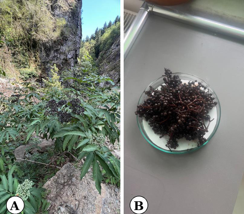
For the preparation of the aqueous extract, a traditional method commonly used in folk practices was adopted. Accordingly, freshly collected fruits were carefully washed with tap water and left to drain overnight to remove excess moisture. Then, 40 grams of fresh fruit were ground with 2000 mL of tap water using a laboratory mill until a homogeneous mixture was obtained. The resulting crude extract was first filtered through muslin cloth, followed by filtration using Whatman No. 1 filter paper to obtain a clear aqueous extract.
Antioxidant activity
DPPH radical scavenging activity
The free radical scavenging capacity of the aqueous extract of Sambucus ebulus fruits was evaluated using the DPPH (2,2-diphenyl-1-picrylhydrazyl) assay, based on the procedure described by Molyneux (2004) with minor modifications. Briefly, 0.75 mL of the extract at various concentrations was mixed with 0.75 mL of 0.1 mM DPPH solution prepared in methanol. The mixture was vortexed and then incubated in the dark at room temperature for 50 minutes. After incubation, the absorbance was measured at 517 nm using a UV-Vis spectrophotometer. Trolox was used as the standard antioxidant. The scavenging activity was expressed as IC₅₀ (mg/mL), which denotes the concentration of extract required to inhibit 50% of the DPPH radicals.
Ferric reducing antioxidant power (FRAP)
The ferric ion reducing capacity of the aqueous extract obtained from Sambucus ebulus fruits was determined based on the method originally described by Benzie and Strain (1996) and further refined by Prior et al. (2005). This assay is based on the principle that, under acidic conditions, antioxidants present in the extract reduce the ferric-tripyridyltriazine (Fe³⁺-TPTZ) complex to its ferrous form (Fe²⁺-TPTZ), resulting in the formation of an intense blue-colored complex. The intensity of the blue color, which directly correlates with the sample’s antioxidant reducing power, was measured spectrophotometrically at 593 nm. The FRAP values were expressed as milligrams of quercetin equivalent per gram of sample (mg QE/g), indicating the extract’s potential to reduce ferric ions.
Total flavonoid content (TFC)
The total flavonoid content in the aqueous extract of Sambucus ebulus fruits was quantified using a modified protocol adapted from Zhishen et al. (1999). This colorimetric method is based on the formation of flavonoid–aluminum chloride complexes. In particular, aluminum ions interact with the C-4 carbonyl and C-3 or C-5 hydroxyl groups of flavones and flavonols, forming stable complexes. Additionally, aluminum chloride may bind to ortho-dihydroxyl substitutions on the A or B rings, though these interactions are generally weaker. Quercetin was used as the reference compound for calibration, with standard solutions ranging from 0.03125 to 1.0 mg/mL. The absorbance of the resulting complexes was measured spectrophotometrically, and the total flavonoid content was expressed as milligrams of quercetin equivalent per gram of sample (mg QE/g).
High-performance liquid chromatography (HPLC) profiling of phenolic compounds
To achieve a comprehensive analysis of structurally diverse phenolic compounds, two different HPLC protocols were utilized, both employing the ACE 5 C18 column (250 × 4.6 mm, i.d.) to maintain consistency in stationary phase selectivity.
Method 1 was tailored to detect common phenolic acids and flavonols. The chromatographic separation employed a binary mobile phase system comprising (A) acetonitrile and (B) 1.5% aqueous acetic acid. The gradient began at 15% A and 85% B, progressively reaching 40% A and 60% B within 29 minutes. The chromatographic system included a 1260 DAD WR detector set at 250, 270, and 320 nm; a 1260 Quaternary Pump operating at a flow rate of 0.7 mL/min; a 1260 Vialsampler administering 10 μL injections; and a G7116A column oven maintained at 35 °C (Table 1).
| Parameter | Method 1 | Method 2 |
|---|---|---|
| Mobile Phase A | Acetonitrile | Methanol |
| Mobile Phase B | 1.5% Acetic Acid | 1.5% Acetic Acid |
| Gradient Start | 15% A / 85% B | 10% A / 90% B |
| Gradient End | 40% A / 60% B (at 29 min) | 90% A / 10% B (40–53 min) |
| Run Time (min) | 29 | 53 |
| Detection Wavelengths (nm) | 250, 270, 320 | 280, 290, 320, 370, 535 |
| Flow Rate (mL/min) | 0.7 | 0.7 |
| Injection Volume (µL) | 10 | 10 |
| Column Temperature (°C) | 35 | 35 |
Method 2 was configured to enhance the separation of phenolic compounds with extended conjugated systems or differing absorption profiles. It utilized a mobile phase composed of (A) methanol and (B) 1.5% aqueous acetic acid. The gradient began at 10% A and 90% B, shifted to 40% A and 60% B by 29 minutes, continued with 60% A and 40% B from 29 to 40 minutes, and ended with 90% A and 10% B from 40 to 53 minutes. Detection wavelengths were set at 280, 290, 320, 370, and 535 nm. Flow rate, injection volume, and column temperature were kept identical to Method 1 (Table 1).
This two-method approach improved analytical reliability and allowed for a broader characterization of the phenolic profile by addressing the chemical heterogeneity of the target compounds.
HS-SPME-GC-MS analysis of volatile compounds
Volatile compound analysis was performed on the aqueous extract prepared from the plant material, which was transferred into headspace vials and tightly sealed with silicone/PTFE septa prior to HS-SPME. The analysis utilized the headspace-solid phase microextraction (HS-SPME) technique, employing an autosampler (PAL RSI, PAL System, Switzerland) equipped with a fiber coated with a composite sorbent layer of divinylbenzene/carbon wide range/polydimethylsiloxane (DVB/C-WR/PDMS, 80 μm).
Samples were pre-equilibrated by incubation at 50 °C for 10 minutes prior to fiber exposure to the headspace, followed by a 10-minute extraction period. Thermal desorption of analytes was achieved by directly inserting the fiber into the GC injection port under splitless mode and maintaining it for 10 minutes. This solvent-free approach allowed efficient capture and transfer of volatile molecules for chromatographic separation.
Gas chromatography was performed using an HP-5MS capillary column (30 m × 0.25 mm, 0.25 μm; Agilent Technologies) with helium as the carrier gas (1 mL/min). The injector operated in splitless mode. The oven temperature was programmed to begin at 50 °C (held for 5 min), then ramped at 3 °C/min to 220 °C, followed by a final isothermal step of 5 minutes. Ionization was conducted via electron impact (EI) at 70 eV, and the mass spectrometer acquired data over an m/z range of 30–500, with a scan rate of 3.1 scans per second.
Compound identification was based on comparison with entries in the NIST 14 (2014) spectral library (National Institute of Standards and Technology, Gaithersburg, MD, USA), with a match quality threshold of ≥85%. Retention indices (RI) were calculated and compared against established literature values for confirmation. Data acquisition and peak deconvolution were processed using Agilent’s MassHunter Qualitative Analysis Workflows. Quantification of volatiles was performed by calculating the percentage of each peak area relative to the total ion chromatogram. Compound identifications were additionally verified by matching retention indices and spectral patterns with those of n-alkane standards.
Experimental design and application procedure
The meristematic cells of Allium cepa (onion, 2n = 16) roots were employed as a biological test system to evaluate morphological and structural alterations in the genetic material and to calculate mitotic indices. Prior to the experiment, the outer scales of the bulbs were carefully removed without damaging the root primordia. The bulbs were then placed in tap water at 22 °C in a dark environment to induce root germination.
For each treatment group, 10 bulbs were initially germinated. Once the root length reached 2–3 cm, five healthy bulbs exhibiting uniform root growth were randomly selected per group. These bulbs were exposed to aqueous extract solutions at different concentrations for 48 hours: 1%, 5%, 10%, 20%, and 50% (v/v, extract/tap water). In addition to the treatment groups, tap water was used as a negative control and a 680 mg/L zinc oxide (ZnO) solution served as the positive control. The ZnO concentration was selected based on previous studies demonstrating its cytotoxic and genotoxic effects in the A. cepa model (Kumari et al. 2011; Ghosh et al. 2016; Debnath et al. 2020). The 48-hour exposure period was determined based on the duration of the mitotic cycle in A. cepa (Boros and Ostafe 2020; Gupta and Kumar 2025). Following this exposure period, both morphological assessments for root growth inhibition and cytogenetic analyses were conducted to investigate genotoxicity indicators in detail.
Assessment of root growth inhibition
To determine the potential inhibitory effects of Sambucus ebulus fruit extract on the primary root development of Allium cepa, root length measurements were carried out after 48 hours of treatment. From each group – including the negative control (tap water), the positive control (680 mg/L ZnO solution), and the extract treatments (1%, 5%, 10%, 20%, and 50% v/v) – a total of 50 roots were randomly selected, with 10 roots taken from each of five bulbs. The root lengths were measured in millimeters and expressed as mean ± standard deviation (SD).
The percentage of root growth inhibition was calculated using the following formula:
Root Growth Inhibition (%) = (Mean root length of control group − Mean root length of treatment group) / Mean root length of control group × 100
This approach enabled a quantitative comparison of inhibitory effects across all treatment groups and revealed the dose-dependent biological response of A. cepa roots to the aqueous extract.
Cytogenetic evaluation
At the end of the treatment period, one root tip was randomly selected from each of the five bulbs in each group, resulting in five slides per treatment group. Root tips approximately 0.5–1 cm in length were excised and fixed in a 3:1 solution of 3% ethanol and 1% acetic acid for 24 hours. The fixed material was then hydrolyzed in 1 N HCl at 60 °C for 5 minutes and subsequently stained with Schiff’s reagent to prepare it for cytogenetic analysis.
Microscopic examination was performed to evaluate three major cytogenetic parameters for each group: mitotic index (MI), micronucleus (MN) frequency in interphase cells, and chromosomal aberrations (CA) observed in dividing cells. These parameters were quantitatively assessed to determine the potential cytotoxic and genotoxic effects of the extract on cell division and genetic material, enabling comparative evaluation across the different concentration groups.
Statistical analysis
All antioxidant assays and Allium cepa-based cytogenetic evaluations were carried out in three independent replicates (n = 3). Results are expressed as mean ± standard deviation (SD). Statistical analyses were performed using SPSS software (version 22.0; IBM Corp., Armonk, NY, USA). To determine statistically significant differences among treatment groups, one-way analysis of variance (ANOVA) was applied. When significant differences were observed, Dunnett’s post hoc test was used to identify the groups differing from the control. Prior to the ANOVA, data were assessed for normality using the Kolmogorov–Smirnov test and for homogeneity of variances using Levene’s test. These tests verified that the assumptions for parametric analysis were met. Statistical significance was accepted at p < 0.05.
Results and discussion
Allium species are widely recognized as reliable biological model systems commonly used to evaluate the cytotoxic and genotoxic effects of plant-derived substances (Tedesco and Laughinghouse 2012, Bonciu et al. 2018; Sarac et al. 2019). In traditional medicine, plants are generally utilized in their entirety or with specific parts (such as leaves, roots, stems) without separation. Therefore, it is of great importance to evaluate the toxicological profiles of herbal products in a holistic manner. Moreover, it has been shown that the bioactive compounds in plants can exert stronger and synergistic effects when interacting with each other rather than acting individually (Tallarida 2011). This suggests that the biological efficacy of plant-based treatments often arises from compound–compound interactions (Rajčević et al. 2022).
In this study, the total flavonoid content (1.008 ± 0.02 mg QE/g), FRAP value (7.045 ± 0.08 mg TE/g), and DPPH radical scavenging activity (0.008 ± 0.30 mg/mL) of the fruit extract of Sambucus ebulus were measured (Table 2). Although the DPPH value was lower than the Trolox standard (0.0033 ± 0.00 mg/mL), the obtained IC₅₀ value is consistent with those reported for aqueous Sambucus species in the literature (Pietta et al. 1998; Rodino et al. 2015). Considering that the aqueous extract is aligned with traditional methods of use (infusion, decoction), the values obtained can be considered biologically relevant. However, studies have reported that methanolic and ethanolic extracts of S. ebulus exhibit higher antioxidant activity in DPPH and FRAP assays (Pietta et al. 1998; Meriç et al. 2014; Rodino et al. 2015). In the studies conducted by ; Pietta et al. (1998) and Rodino et al. (2015), DPPH inhibition levels of methanolic extracts were found to reach 80–90%, while aqueous extracts generally ranged between 40–60%. This has been attributed to the better solubility of polyphenols and flavonoids in organic solvents. Similar trends have also been reported for S. nigra and S. canadensis species (Sidor and Gramza-Michałowska 2015; Januškevičė et al. 2025). Ethanolic and acetonic extracts obtained from the fruits and flowers of S. nigra were shown to be associated with high phenolic content and strong antioxidant activity (Sidor and Gramza-Michałowska 2015; Januškevičė et al. 2025). Although interspecies differences are to be considered, the current findings support that S. ebulus is a strong natural antioxidant source.
| Parameter | Value (Mean ± SD) |
|---|---|
| Total Flavonoid Content (mg QE/g) | 1.008 ± 0.02 |
| Ferric Reducing Antioxidant Power (FRAP) (mg QE/g) | 7.045 ± 0.08 |
| DPPH Radical Scavenging Activity (IC50 Inhibition-mg/ml) | 0.008 ± 0.30 |
| Standard (IC50 DPPH Inhibition (mg/ml) | 0.0033±0.00 |
It is well established that antioxidants not only have chemical potential but also exert cytogenetic effects at the cellular level. Reactive oxygen species (ROS) can lead to DNA damage, chromosomal abnormalities, and mitotic disturbances, while antioxidants may prevent such harmful effects (Meriç et al., 2014; Sidor and Gramza-Michałowska 2015; Kayıran et al. 2022; Januškevičė et al. 2025). Therefore, it is presumed that the antioxidant capacity of the aqueous extract used in this study may be effective in reducing chromosomal damage or preserving mitotic activity. Supporting this assumption, Meriç et al. (2014) reported that the high phenolic content and DPPH activity in the methanolic extract of S. ebulus were associated with non-cytotoxic but potentially anticarcinogenic effects. Similarly, Rodino et al. (2015) demonstrated strong anti-oxidative stress effects with high FRAP (1725.5 ± 9.8 μmol Fe²⁺/g) and DPPH (85.3 ± 0.2%) values in S. ebulus fruit extract.
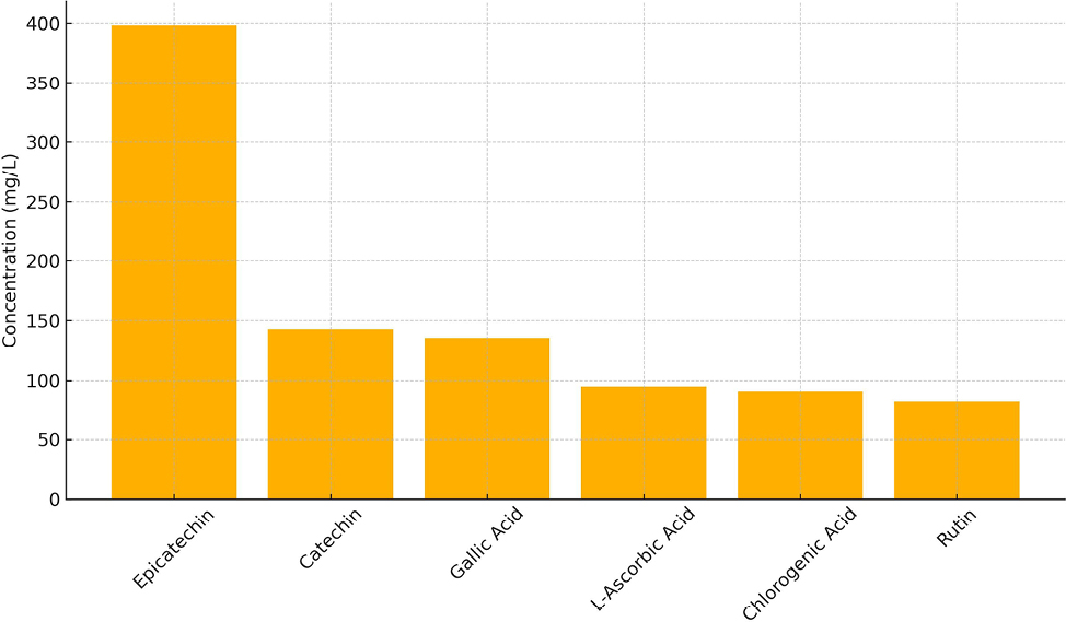
HPLC analyses revealed that the extract contains significant phenolic compounds such as epicatechin (398.29 mg/L), catechin (142.86 mg/L), gallic acid (135.4 mg/L), chlorogenic acid (89.58 mg/L), L-ascorbic acid (94.98 mg/L), and rutin (81.43 mg/L) (Table 3, Figure 2). According to the literature, flavonoids such as epicatechin and catechin are known to be involved in processes related to cell cycle regulation and the enhancement of mitotic activity (Alimullah et al. 2025; Aljuhaimi et al. 2025; Sancer et al. 2025; Uslu et al. 2025;). Therefore, it is plausible that these compounds may exert beneficial effects on cytogenetic parameters. Phenolic acids like gallic acid and chlorogenic acid have also been identified as bioactive molecules with potential roles in reducing DNA damage and maintaining chromosomal integrity. In this context, Amić et al. (2025) and Signorini et al. (2025) have reported that these compounds might contribute to the preservation of genetic material by mitigating oxidative DNA damage. Furthermore, L-ascorbic acid is not only a potent antioxidant but also has been associated with regulatory functions in the cell cycle Shams (Shams El Dine et al. 2025). The presence of this compound in the extract may help explain the observed regulatory effects on mitotic activity. The coexistence of these compounds is likely to enhance the overall antioxidant capacity of the extract and may promote synergistic interactions, further supporting its biological efficacy (Tallarida 2011).
| No | Compounds | (mg/L) |
|---|---|---|
| Vitamin | ||
| 1 | L-Ascorbic acid | 94.98 |
| Phenolics | ||
| 2 | Gallic acid | 135.4 |
| 3 | 3,4-Dihydroxy benzoic acid | 53.1 |
| 4 | Vanillic acid | N/D |
| 5 | Syringic acid | N/D |
| 6 | p-Coumarıc Acid | N/D |
| 7 | Trans-Caffeic acid | 46.69 |
| 8 | Ferulic acid | |
| 9 | Rosmarinic acid | 3.89 |
| 10 | Pyrogallol | 6.48 |
| 11 | Chlorogenic acid | 89.58 |
| 12 | Resveratrol | N/D |
| 13 | Oleuropein | N/D |
| Flavonoids | ||
| 14 | (+)-Catechin | 142.86 |
| 15 | (-)-Epicatechin | 398.29 |
| 16 | Rutin | 0.5 |
| 17 | Myricetin | N/D |
| 18 | Quercetin | N/D |
| 19 | Apigenin | N/D |
| 20 | Cyanidin chloride | N/D |
| 21 | Hesperitin | N/D |
| 22 | Kaempferol | N/D |
| 23 | Baicalien | N/D |
| 24 | Chrysin | N/D |
According to GC-MS analysis, more than 95% of the volatile profile consisted of compounds such as ethyl isovalerate, methyl isovalerate, and trans-β-ocimene (Table 4). These volatiles have been linked in the literature to antioxidant, anti-inflammatory, and membrane-regulating properties (Calva et al. 2025; Jaishi et al. 2025; Otto et al. 2025). In addition, o-cymene and butanoic acid derivatives have been reported to enhance bioactivity through interactions with cellular membranes (Dorman and Deans 2000). This chemical diversity suggests that the extract may possess a biological potential capable of supporting cytogenetic activity (Rajčević et al. 2022).
| No | RT (min) | RI | Name of the compound | Content [%] |
|---|---|---|---|---|
| 1 | 3.891 | 650 | Butanal, 3-methyl- | 4.162 |
| 2 | 4.329 | 716 | Pentanal | 1.285 |
| 3 | 5.899 | 790 | Methyl isovalerate | 24.848 |
| 4 | 6.501 | 812 | Hexanal | 3.611 |
| 5 | 6.775 | 821 | Butanoic acid, 3-methyl-, ethyl ester | 10.544 |
| 6 | 7.095 | 830 | Butanoic acid, 3-methyl- | 4.775 |
| 7 | 8.209 | 827 | Isovaleric acid, ethyl ester | 25.277 |
| 8 | 14.344 | 1016 | o-Cymene | 7.486 |
| 9 | 14.575 | 1021 | trans-.beta.-Ocimene | 14.421 |
| 10 | 16.329 | 1059 | gamma.-Terpinene | 3.591 |
The dose-dependent cytogenetic effects of the extract were revealed through analyses of root elongation, mitotic index (MI), chromosomal abnormalities, and micronucleus formation. The effect of the aqueous extract of Sambucus ebulus on root elongation was observed in the negative control group, where the highest mean root length (61.84 ± 7.96 mm) was recorded. This result indicates that mitotic activity proceeded normally and that there was no impairment in cellular proliferation. In contrast, in the positive control group (ZnO treatment), a significant reduction in root length (10.02 ± 5.53 mm) was detected, confirming the test’s sensitivity to toxic agents with an inhibition rate of 83.78% (Table 5, Figure 3).
| Treatments | Examined root number | Root length (mm) (Mean±SD) | Inhibition (%) | ||||||||||||||||
|---|---|---|---|---|---|---|---|---|---|---|---|---|---|---|---|---|---|---|---|
| Negative control | 50 | 61.84±7.96 | - | ||||||||||||||||
| Positive control | 50 | 10.02±5.53* | 83.78 | ||||||||||||||||
| 1% | 50 | 41.93±6.00 | 32.18 | ||||||||||||||||
| 5% | 50 | 46.76±6.09 | 27.61 | ||||||||||||||||
| 10% | 50 | 49.31±5.89 | 20.27 | ||||||||||||||||
| 20% | 50 | 31.29±6.34* | 23.23 | ||||||||||||||||
| 50% | 50 | 23.92±5.26* | 61.32 | ||||||||||||||||
| *Significant at p<0.05. | |||||||||||||||||||
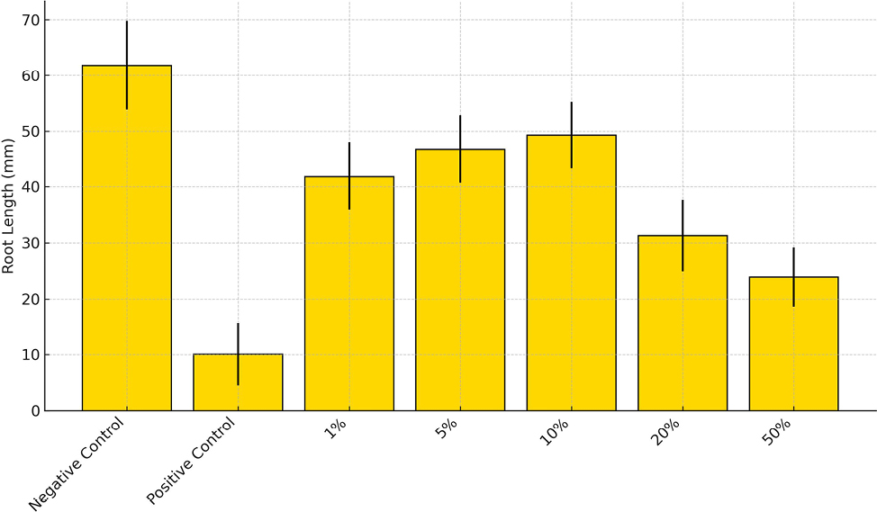
Among the extract-treated groups, a dose-dependent inhibition of root growth was observed as extract concentration increased. Notably, 50% (23.92 ± 5.26 mm) and 20% (31.29 ± 6.34 mm) concentrations resulted in significant reductions in root length with inhibition rates of 61.32% and 23.23%, respectively (Table 5, Figure 3). These effects were statistically significant compared to the negative control, suggesting that the extract contains bioactive compounds capable of affecting cell division and elongation processes (Chauhan et al. 1999; Murthy et al. 2011).
However, the relatively low inhibition rates observed at moderate concentrations (1% to 10%) and the nonlinear response between 10% and 20% suggest that some compounds may exhibit hormetic effects at low doses or may activate non-cytotoxic mechanisms (Chauhan et al., 1999; Murthy et al. 2011). Previous studies have also reported that plant extracts rich in polyphenols and terpenoids may inhibit root growth in A. cepa by disrupting mitotic spindle formation and interfering with cell wall synthesis (Leme and Marin-Morales 2009; Celik 2012).
The effect on mitotic activity was assessed by considering MI values and the distribution of cells at different mitotic stages in A. cepa root meristem tissue. The high MI value observed in the negative control group (7.71 ± 1.07%) indicates that the mitotic process functioned physiologically without disruption. In contrast, the MI in the ZnO-treated positive control group decreased significantly to 2.24 ± 0.04% (p < 0.05), confirming that ZnO inhibits mitosis and that the Allium test is a reliable model for detecting cytotoxic effects (Table 6).
| Treatments | Examined Cell Number | MI±SD | Total number of cells at prophase | Total number of cells at metaphase | Total number of cells at anaphase | Total number of cells at telophase | |||||||||||||
|---|---|---|---|---|---|---|---|---|---|---|---|---|---|---|---|---|---|---|---|
| Negative control | 5000 | 7.71±1.07 | 440 | 200 | 60 | 110 | |||||||||||||
| Positive control | 5000 | 2.24±0.04* | 170 | 120 | 50 | 40 | |||||||||||||
| 1% | 5000 | 4.98±0.50 | 360 | 70 | 60 | 30 | |||||||||||||
| 5% | 5000 | 5.26±0.90 | 430 | 60 | 70 | 30 | |||||||||||||
| 10% | 5000 | 6.55±3.16 | 330 | 180 | 70 | 80 | |||||||||||||
| 20% | 5000 | 2.56±0.39* | 200 | 90 | 60 | 20 | |||||||||||||
| 50% | 5000 | 2.57±0.10* | 170 | 140 | 70 | 2 | |||||||||||||
| *Significant at p<0.05. | |||||||||||||||||||
In the extract-treated groups, MI values varied depending on the applied concentration. At low concentrations (1%, 5%, and 10%), MI was determined as 4.98 ± 0.50%, 5.26 ± 0.90%, and 6.55 ± 3.16%, respectively (Table 6). These values were significantly higher than those of the positive control group, suggesting that these doses did not suppress mitotic activity and may even support limited proliferation. This aligns with the concept of hormesis, which posits that certain natural compounds can stimulate cellular responses at low doses (Calabrese and Baldwin 2003). However, at higher concentrations (20%: 2.56 ± 0.39%; 50%: 2.57 ± 0.10%), MI values dropped to levels comparable to the positive control, indicating that these doses markedly suppressed cell division.
MI values changed inversely with concentration, with lower doses supporting mitotic activity, while significant reductions in MI were observed in the 20% and 50% groups. This decline may be explained by the ability of phenolic acids and volatile terpenes to inhibit mitosis at the G2/M phase (Akaneme and Amaefule 2012; Yıldız et al. 2025).
Chromosomal abnormalities, particularly C-mitosis, chromosome stickiness, and bridge formation, increased markedly at higher doses. Similarly, micronucleus frequencies showed significant increases at 20% and 50% concentrations, while these effects remained minimal in the 1–10% groups. These findings indicate that genotoxic effects are dose-threshold dependent and that cell division is not impaired at low concentrations (Calabrese and Baldwin, 2003; Ždralović et al. 2019; Smirnova and Korovkina 2021).
Structural anomalies commonly observed in the positive control group – particularly C-mitosis, chromosome stickiness, and chromatid bridges – were also detected in the high-dose extract treatment groups. For example, in the 50% extract group, the frequency of C-mitosis (7.33 ± 0.00), bridge formation (4.00 ± 1.41), and chromosome stickiness (3.17 ± 0.32) were considerably high (Table 7, Figure 4). These anomalies are associated with disruptions in the formation of mitotic spindle fibers during the metaphase–anaphase transition (Yüzbaşıoğlu et al. 2003; Smirnova and Korovkina 2021).
| Treatments | Disturbed prophase | Chromosome stickness | C-mitosis | Chromatid bridge | Laggard chromosome | Vagrant chromosome | Total abberant cells | ||||||||||||
|---|---|---|---|---|---|---|---|---|---|---|---|---|---|---|---|---|---|---|---|
| Negative control | - | 0.33±0.52 | - | 0.67±0.02 | - | - | 1.00±0.21 | ||||||||||||
| Positive control | 4.00±0.41 | 4.83±1.06 | 9.00±0.00 | 6.67±0.05 | 0.50±0.06 | 2.50±0.71 | 27.50±0.71* | ||||||||||||
| 1% | - | 0.50±0.14 | 1.00±0.03 | 0.50±0.51 | - | 1.00±0.31 | 3.00±1.1 | ||||||||||||
| 5% | 0.67±0.01 | 0.33±0.02 | - | 0.50±0.01 | - | 1.50±0.11 | 3.00±0.7 | ||||||||||||
| 10% | - | 0.78±1.20 | 0.33±0.58 | 1.56±0.02 | - | - | 2.67±0.1 | ||||||||||||
| 20% | - | 4.00±0.43 | 2.50±0.71 | 3.50±0.60 | - | 1.50±0.41 | 11.50±0.71* | ||||||||||||
| 50% | 2.50±0.71 | 3.17±0.32 | 7.33±0.00 | 4.00±1.41 | - | 4.00±1.01 | 21.00±1.02* | ||||||||||||
| *Significant at p<0.05. | |||||||||||||||||||
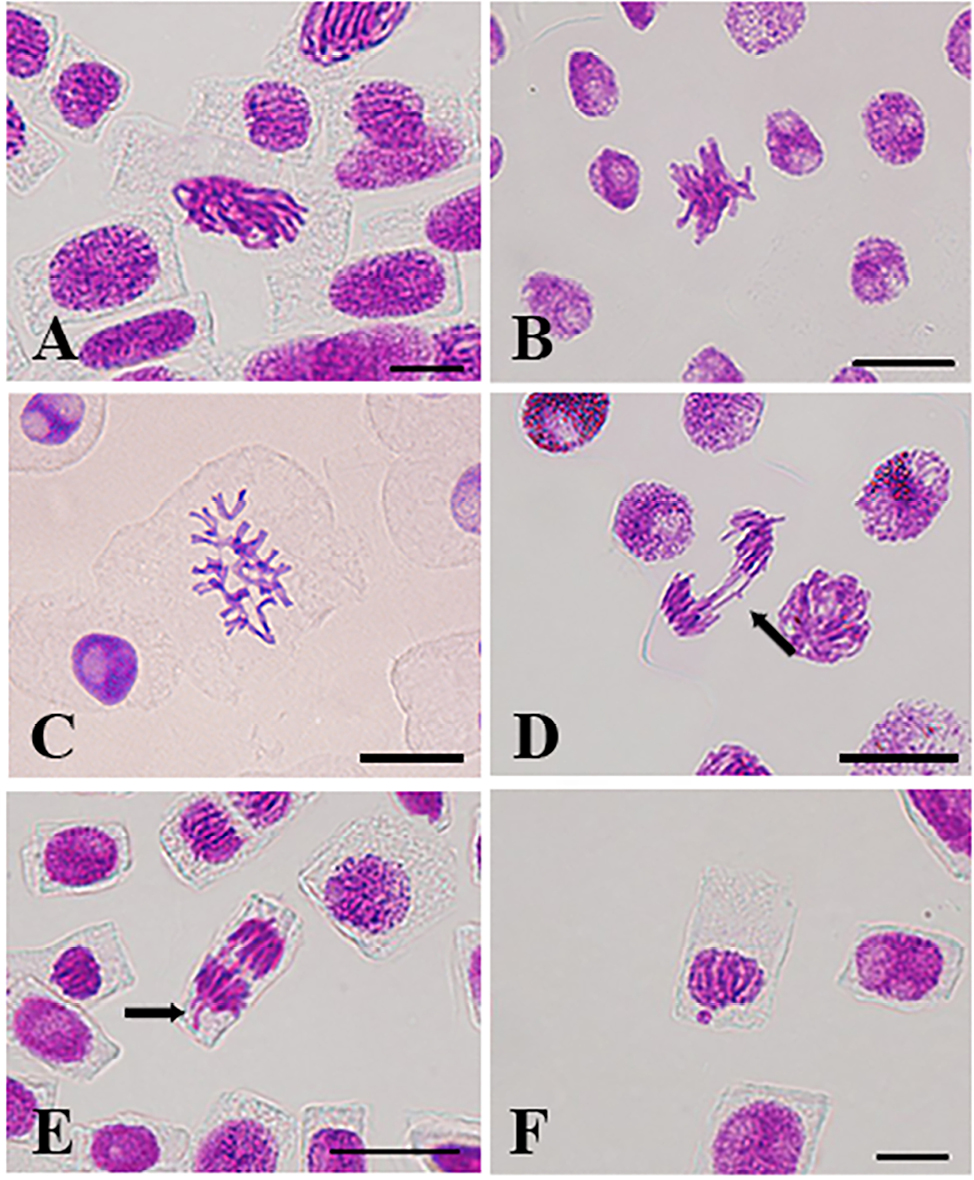
In contrast, a lower number of abnormal cells were observed in the extract treatments administered at low and moderate concentrations, and structural abnormalities generally remained at a mild level. This dose-dependent trend suggests that increasing extract concentration enhances genotoxic stress and exacerbates chromosomal damage. Particularly in the 20% and 50% groups, the total numbers of abnormal cells were recorded as 11.50 ± 0.71 and 21.00 ± 1.02, respectively (Table 7).
The formation of C-mitosis indicates the presence of agents that disrupt spindle fiber development, whereas chromosome stickiness may hinder the proper segregation of chromosomes, potentially leading to mitotic arrest (Asita et al. 2021; Pharmawati et al. 2022). Bridges and lagging chromosomes are mitotic anomalies resulting from improper chromosome separation toward the poles and are commonly associated with DNA strand breaks or replication errors (Leme and Marin-Morales 2009; Asita et al. 2021).
The obtained micronucleus (MN) data indicate that the Sambucus ebulus fruit extract may exhibit genotoxic potential at high concentrations, whereas its effects remain minimal at lower concentrations (Table 8). Statistically significant increases in MN frequency were observed particularly in the 20% and 50% treatment groups, indicating that these doses may induce DNA damage and genomic instability. However, in the 1% and 5% extract groups, MN frequencies remained similar to those of the negative control, and no micronucleus formation was detected at the 10% concentration (Table 8). These findings point to the possibility of a hormetic response, suggesting that the extract does not impair cytogenetic stability below a certain threshold concentration (Calabrese and Baldwin, 2003).
| Treatments | Examined Cell Number | Micronucleus (%) | |||||||||||||||||
|---|---|---|---|---|---|---|---|---|---|---|---|---|---|---|---|---|---|---|---|
| Negative control | 5000 | - | |||||||||||||||||
| Positive control | 5000 | 1.35±0.02* | |||||||||||||||||
| 1% | 5000 | 0.05±0.01 | |||||||||||||||||
| 5% | 5000 | 0.05±0.01 | |||||||||||||||||
| 10% | 5000 | - | |||||||||||||||||
| 20% | 5000 | 0.45±0.07* | |||||||||||||||||
| 50% | 5000 | 1.05±0.02* | |||||||||||||||||
| *Significant at p<0.05. | |||||||||||||||||||
Conclusion
This study demonstrates that the aqueous extract obtained from the fruits of Sambucus ebulus exhibits notable antioxidant and cytogenetic effects through its bioactive compounds. While applications at low concentrations displayed supportive effects on cellular antioxidant defense and preservative effects on mitotic activity, higher concentrations led to suppression of the cell cycle and structural damage to the genetic material. These findings indicate that compounds such as phenolic acids and volatile terpenes may elicit different biological responses depending on their concentration and interaction level (Sultan and Çelik 2009; Smirnova and Korovkina 2021; Pharmawati et al. 2022). Considering that S. ebulus is widely used in traditional medicine for immunomodulatory, anti-inflammatory, and antiviral purposes, the aim of this study is not to label the plant as harmful, but rather to draw attention to the potential cytogenetic risks associated with concentrations exceeding certain threshold doses. In particular, concentrations at or below 10%, where no micronucleus formation was observed, provide preliminary data that could serve in defining safe usage ranges for the aqueous extract of Sambucus ebulus. The results reveal that S. ebulus, with its rich profile of bioactive compounds, has the potential to modulate cell proliferation and genetic stability; they also emphasize the necessity of carefully determining dose ranges for safe and effective use.
References
Akaneme FI, Amaefule CC. 2012. Evaluation of the cytotoxicity and genotoxicity of aqueous leaf extracts of Azadirachta indica A. Juss using the Allium test. Journal of Medicinal Plants Research. 6(22):3898–3907.
Alimullah M, Jahan I, Rahman MM, Khan F, Ahmed KS, Hossain MH, Subhan N. 2025. Camellia sinensis powder rich in epicatechin and polyphenols attenuates isoprenaline-induced cardiac injury by activating the Nrf2–HO1 antioxidant pathway in rats. Scientific Reports. 15:20859. https://doi.org/10.1038/s41598-025-08720-w
Aljuhaimi F, Uslu N, Özcan MM, Ahmed IAM, Walayat N. 2025. Investigation of changes in the bioactive properties and phenolic compositions of different layers of the leek plant. Acta Scientiarum Polonorum Technologia Alimentaria. 24:447–456.
Amić A, Milanović Ž, Cagardová DM. 2025. Protective roles of α-tocopherol, α-CEHC, and gallic acid against CO₃•⁻-induced DNA damage and their effect on OGG1 and pro-oxidant enzymes. Journal of Molecular Liquids. 438:128847. https://doi.org/10.1016/j.molliq.2025.128847
Asita AO, Maga-ma S, Moahloli TM, Baholo S. 2021. Evaluation of extracts of wild Cannabis sativa L. for genotoxicity and phytochemical composition. Caryologia. 74(1):135–150.
Barak TH, Celep E, İnan Y, Yeşilada E. 2020. In vitro human digestion simulation of the bioavailability and antioxidant activity of phenolics from Sambucus ebulus L. fruit extracts. Food Bioscience. 37:100711. https://doi.org/10.1016/j.fbio.2020.100711
Benzie IFF, Strain JJ. 1996. The ferric reducing ability of plasma (FRAP) as a measure of “antioxidant power”: the FRAP assay. Analytical Biochemistry. 239:70–76. https://doi.org/10.1006/abio.1996.0292
Boros BV, Ostafe V. 2020. Evaluation of ecotoxicology assessment methods of nanomaterials and their effects. Nanomaterials. 10:610. https://doi.org/10.3390/nano10040610
Brasil. Ministério da Saúde. Secretaria de Ciência, Tecnologia e Insumos Estratégicos. 2006. Política Nacional de Plantas Medicinais e Fitoterápicos. Brasília (Brazil): Editora MS.
Bonciu E, Firbas P, Fontanetti CS, Wusheng J, Karaismailoğlu MC, Liu D, et al. 2018. An evaluation for the standardization of the Allium cepa test as cytotoxicity and genotoxicity assay. Caryologia. 71(3):191–209. https://doi.org/10.1080/00087114.2018.1503496
Calabrese EJ, Baldwin LA. 2003. Hormesis: the dose-response revolution. Annual Review of Pharmacology and Toxicology. 43(1):175–197. https://doi.org/10.1146/annurev.pharmtox.43.100901.140223
Calva J, Cuenca MB, León A, Benítez Á. 2025. Chemical composition, acetylcholinesterase-inhibitory potential and antioxidant activity of essential oils from three populations of Parthenium hysterophorus L. in Ecuador. Molecules. 30:2712. https://doi.org/10.3390/molecules30132712
Chauhan LKS, Saxena PN, Gupta SK. 1999. Cytogenetic effects of cypermethrin and fenvalerate on the root meristem cells of Allium cepa. Environmental and Experimental Botany. 42:181–189. https://doi.org/10.1016/S0098-8472(99)00033-7
Celik TA. 2012. Potential genotoxic and cytotoxic effects of plant extracts. In: A compendium of essays on alternative therapy. p. 233–250.
Davis PH. 1970. Flora of Turkey and the East Aegean Islands. Edinburgh (UK): Edinburgh University Press.
Debnath P, Mondal A, Sen K, Mishra D, Mondal NK. 2020. Genotoxicity study of nano Al₂O₃, TiO₂ and ZnO along with UV-B exposure: an Allium cepa root tip assay. Science of the Total Environment. 713:136592. https://doi.org/10.1016/j.scitotenv.2020.136592
Dorman HD, Deans SG. 2000. Antimicrobial agents from plants: antibacterial activity of plant volatile oils. Journal of Applied Microbiology. 88(2):308–316.
Shams El Dine RS, Youseef HT, Awaad AK, Hammoury SI, Mohamed EI. 2025. The potentially therapeutic effects of ascorbic acid in different cell lines in an attempt to reduce the risk of radiation therapy. Scientific Reports. 15:15077. https://doi.org/10.1038/s41598-025-96697-x
Ghosh M, Jana A, Sinha S, Jothiramajayam M, Nag A, Chakraborty A, et al. 2016. Effects of ZnO nanoparticles in plants: cytotoxicity, genotoxicity, deregulation of antioxidant defenses, and cell-cycle arrest. Mutation Research/Genetic Toxicology and Environmental Mutagenesis. 807:25–32. https://doi.org/10.1016/j.mrgentox.2016.07.006
Gupta S, Kumar A. 2025. Cytogenetic analysis of mitotic cell cycle phases in Allium cepa root meristem: a model for chromosomal behaviour and environmental toxicity assessment. International Internal Medicine Journal. 3:1–5.
Hister CAL, Boligon AA, Laughinghouse HD IV, Tedesco SB. 2017. Determination of phenolic compounds and assessment of the genotoxic and proliferative potential of Psidium cattleianum Sabine (Myrtaceae) fruits. Caryologia. 70:350–356. https://doi.org/10.1080/00087114.2017.1360571
Jaishi DR, Ojha I, Ojha SR, Pal P, Thalal B, Chaudhary PN. 2025. Extraction, chemical composition, antioxidant, and antibacterial activities of essential oil using aerial parts of Artemisia indica. Journal of the Nepal Chemical Society. 45:34–44. https://doi.org/10.3126/jncs.v45i2.82974
Januškevičė V, Saunoriūtė S, Ondrášek I, Petrikaitė V, Štreimikytė P, Liaudanskas M, Česonienė L. 2025. Biochemical diversity and anticancer potential of phenolic compounds in Sambucus nigra L. cultivars. Applied Food Research. 5:101404. https://doi.org/10.1016/j.afres.2025.101404
Kayıran SD, Özkan EE, Kara EM, Tarı Ö, Nenni M. 2022. Determination of the chemical composition and antioxidant potential of Sambucus ebulus L. (dwarf elder) fruit extracts and investigation of antimicrobial activity on Trichophyton rubrum (Castell.) Sabour and some microorganisms. Istanbul Journal of Pharmacy. 52(2):148–155. https://doi.org/10.26650/IstanbulJPharm.2022.929762
Kumari M, Khan SS, Pakrashi S, Mukherjee A, Chandrasekaran N. 2011. Cytogenetic and genotoxic effects of zinc oxide nanoparticles on root cells of Allium cepa. Journal of Hazardous Materials. 190(1–3):613–621. https://doi.org/10.1016/j.jhazmat.2011.03.095
Leme DM, Marin-Morales MA. 2009. Allium test in monitoring. Mutation Research. 682:71–81. https://doi.org/10.1016/j.mrrev.2009.06.002
Martins ER, Castro DM, Castellani DC, Dias JE. 2003. Plantas medicinais. Viçosa (Brazil): Editora UFV.
Meriç Z, Bitiş L, Tan SB, Turan SÖ, Akbuğa J. 2014. Antioxidant, antimicrobial and anticarcinogenic activities of Sambucus ebulus L. flowers, fruits and leaves. Marmara Pharmaceutical Journal. 18(1):22–25.
Mohan AR, Joseph N. 2024. Cytotoxic assessment of aqueous extracts of Heliotropium keralense Sivar. & Manilal on Allium cepa root tip cells. Caryologia. 77(2):29–36. https://doi.org/10.36253/caryologia-2436
Molyneux P. 2004. The use of the stable free radical diphenylpicrylhydrazyl (DPPH) for estimating antioxidant activity. Songklanakarin Journal of Science and Technology. 26:211–219.
Murthy GS, Francis TP, Singh CR, Nagendra HG, Naik C. 2011. An assay for screening anti-mitotic activity of herbal extracts. Current Science. 100:1399–1404. https://www.jstor.org/stable/24076606
National Institute of Standards and Technology (NIST). 2014. NIST/EPA/NIH mass spectral library. Version 14. Gaithersburg (MD, USA): National Institute of Standards and Technology.
Otto P, Ninkovic V, Meiners T, Pashalidou FG, Fortuna TM, Louis J, Cusumano A. 2025. Multifunctionality of plant VOCs in agroecological systems: perspectives for biological pest control. Entomologia Generalis. https://doi.org/10.1127/entomologia/3506
Pharmawati M, Wirasiti NN, Wrasiati LP. 2022. Genotoxic and antigenotoxic potential of encapsulated Enhalus acoroides (L. f.) Royle leaves extract against nickel nitrate. Caryologia. 75(2):89–99.
Pietta P, Simonetti P, Mauri P. 1998. Antioxidant activity of selected medicinal plants. Journal of Agricultural and Food Chemistry. 46:4487–4490. https://doi.org/10.1021/jf980310p
Prior RL, Wu X, Schaich K. 2005. Standardized methods for the determination of antioxidant capacity. Journal of Agricultural and Food Chemistry. 53:4290–4302. https://doi.org/10.1021/jf0502698
Rajčević N, Bukvički D, Dodoš T, Marin PD. 2022. Interactions between natural products – a review. Metabolites. 12:1256. https://doi.org/10.3390/metabo12121256
Rank J, Nielsen MH. 1994. Evaluation of the Allium anaphase–telophase test in relation to genotoxicity screening of industrial wastewater. Mutation Research. 312:17–24. https://doi.org/10.1016/0165-1161(94)90004-3
Rodino S, Butu A, Petrache P, Butu M, Dinu-Pîrvu CE, Cornea CP. 2015. Evaluation of the antimicrobial and antioxidant activity of Sambucus ebulus extract. Farmacia. 63:751–754.
Sancer O, Tepebaşı MY, Şahin U, İlhan İ. 2025. Uncovering the benefits of epicatechin for oxidative stress in human health. Süleyman Demirel University Journal of Health Sciences. 16:75–81. https://doi.org/10.22312/sdusbed.1539732
Sarac I, Bonciu E, Butnariu M, Petrescu I, Madosa E. 2019. Evaluation of the cytotoxic and genotoxic potential of some heavy metals by use of Allium test. Caryologia. 72(2):37–43.
Shams El Dine RS, Youseef HT, Awaad AK, Hammoury SI, Mohamed EI. 2025. The potentially therapeutic effects of ascorbic acid in different cell lines in an attempt to reduce the risk of radiation therapy. Scientific Reports. 15:15077. https://doi.org/10.1038/s41598-025-96697-x
Sidor A, Gramza-Michałowska A. 2015. Advanced research on the antioxidant and health benefit of elderberry (Sambucus nigra) in food – a review. Journal of Functional Foods. 18:941–958. https://doi.org/10.1016/j.jff.2014.07.012
Signorini C, Corsaro R, Collodel G, Maettner R, Sterzik K, Strehler E, Moretti E. 2025. Addition of chlorogenic acid to human semen: effects on sperm motility, DNA integrity, oxidative stress, and Nrf2 expression. Antioxidants. 14:382. https://doi.org/10.3390/antiox14040382
Smirnova MV, Korovkina AV. 2021. Toxic and genotoxic effects of aqueous extracts of Polygonum weyrichii Fr. Schmidt on the Allium test taken as an example. Caryologia. 74(2):79–88.
Sousa C, Silva R, Costa J, Figueiredo N, Oliveira A, Santos D, Almeida M. 2018. In vivo assessment of cyto/genotoxic, antigenotoxic and antifungal potential of Costus spiralis (Jacq.) Roscoe leaves and stems. Biocatalysis and Agricultural Biotechnology. 14:135–143. https://doi.org/10.1016/j.bcab.2018.02.007
Sultan AÖ, Çelik TA. 2009. Genotoxic and antimutagenic effects of Capparis spinosa L. on the Allium cepa L. root tip meristem cells. Caryologia. 62(2):114–123.
Tallarida RJ. 2011. Quantitative methods for assessing drug synergism. Genes & Cancer. 2(11):1003–1008. https://doi.org/10.1177/194760191244057.
Tedesco SB, Laughinghouse HD IV. 2012. Bioindicator of genotoxicity: the Allium cepa test. In: Srivastava JK, editor. Environmental contamination. Rijeka (Croatia): InTech. p. 137–156. https://doi.org/10.5772/35355
Tedesco M, Kuhn AW, Boligon AA, Laughinghouse HD IV, Athayde ML, Silva ACS, Tedesco SB. 2015. Chromatographic analysis, antiproliferative effect and genotoxicity of aqueous extracts of Citrus sinensis (L.) Osbeck on the Allium cepa L. test system. Bioscience Journal. 31:1213–1221.
Teixeira RO, Camparoto ML, Mantovani MS, Vicentini VEP. 2003. Assessment of two medicinal plants, Psidium guajava L. and Achillea millefolium L., in vitro and in vivo assays. Genetics and Molecular Biology. 26:551–555. https://doi.org/10.1590/S1415-47572003000400021
Trapp KC, Hister CAL, Laughinghouse HD IV, Boligon AA, Tedesco SB. 2020. Determination of phenolic compounds and evaluation of cytotoxicity in Plectranthus barbatus using the Allium cepa test. Caryologia. 73(2):145–153.
Tuna-Gülören Ö, Korkmaz F, Erdir M, Ataşlar E. 2021. Cytotoxic and genotoxic effects of methanol extracts of vegetative parts of some Gypsophila L. species using Allium cepa assay. Caryologia. 74(2):141–148.
Uslu K, Çavuşoğlu K, Yalçın E. 2025. Quantitative phenolic profiling and protective effects of grape seed extract on mancozeb-induced cellular and genetic toxicity. Scientific Reports. 15:36249. https://doi.org/10.1038/s41598-025-20204-5
Yeşilada E, Gürbüz İ, Toker G. 2014. Anti-ulcerogenic activity and isolation of the active principles from Sambucus ebulus L. leaves. Journal of Ethnopharmacology. 153:478–483. https://doi.org/10.1016/j.jep.2014.03.004
Yıldız E, Meredova G, Aksoy H. 2025. The genoprotective role of Laurocerasus officinalis: study on genotoxic/antigenotoxic and cytotoxic/anticytotoxic effects in human lymphocytes. Journal of Food Biochemistry. 2025:7455954. https://doi.org/10.1155/jfbc/7455954
Yüzbaşıoğlu D, Ünal F, Sancak C, Kasap R. 2003. Cytological effects of the herbicide racer “flurochloridone” on Allium cepa. Caryologia. 56(1):97–105.
Ždralović A, Mešić A, Eminović I, Parić A. 2019. Cytotoxic and genotoxic activity of Plantago major L. extracts. Caryologia. 72(3):35–40.
Zhishen J, Mengcheng T, Jianming W. 1999. The determination of flavonoid contents in mulberry and their scavenging effects on superoxide radicals. Food Chemistry. 64:555–559. https://doi.org/10.1016/S0308-8146(98)00102-2