Received: August 13, 2025; Accepted: December 2, 2025; Published: Month 11, 2026
A comprehensive study of UV-B effects on Salvia hispanica L. (Lamiaceae): Genetic variation and biochemical responses in M1 generation
Department of Botany, University of Allahabad, UP-211002 (India) Prayagraj, India
Email: satyap.sp90@gmail.com; kumar_girjesh@yahoo.com
*Corresponding author
Abstract. This study investigated the impact of different durations of UV-B exposure on Salvia hispanica L. The main objective was to assess the genetic and metabolic responses of chia plants, with a focus on morphological changes, phytochemical analysis, and cytological abnormalities. Chia seedlings were exposed to UV-B radiation for different periods, ranging from 0 to 50 minutes, with subsequent one-hour recovery periods at room temperature. The primary finding of this study was that subjecting chia seedlings to a moderate UV-B dose for 20 minutes yielded favorable outcomes. This included a significant increase in leaf area (9.02%) and a 25% increase in plant height compared to the control group, both of which were statistically significant (p<0.05). However, exposure to plant emergence of chlorophyll mutants (Xantha, semi-xantha, and Albina) and tall, bushy plant mutants in the M2 generation. In cytological observation resulted in chromosomal aberrations, such as stickiness, scattering, and unorientation, implying potential genotoxic impacts. Biochemical assessments revealed a significant increase in photosynthetic pigment content, particularly Chl a (12.91%) and Chl b (17.14%), both significantly elevated (p<0.05) compared to the control group. Metabolite analysis through GC-MS uncovered notable variations in fatty acid composition, with higher quantities of alpha-linolenic acid (1.24%), gamma-tocopherol (1.2%), and linolenic acid methyl ester (25.18%) observed in the treatment group exposed to short-wavelength UV-B compared to the control set.. Further research is needed to elucidate the underlying mechanisms and practical applications of these findings.
Keywords: Salvia hispanica L., GC-MS analysis, chromosomal abnormality, UV-B short exposure, Principal Component Analysis (PCA), LD50.
Contents
Experimental design - UV - B treatment
Chlorophyll mutants in response to UV-B exposure
UV-B exposure study through multivariate statistical analysis (MVA) and Pearson correlation matrix
Highlights
– Optimizing UV-B treatment duration is crucial for effective plant treatment and maximizing resourceful metabolite production.
– The study examines the effects of varying UV-B exposure durations on Salvia hispanica L. (chia) plants, focusing on genetics and metabolism.
– Biochemical analyses showed increased photosynthetic pigment content, notably Chl a (12.91%) and Chl b (17.14%), compared to control.
– Exposure to moderate UV-B for 20 minutes led to significant positive changes, including increased leaf area (9.02%) and a 25% taller plant height.
– Controlled 20-minute UV-B exposure led to beneficial shifts in chia plants, affecting both morphology and biochemistry.
Introduction
Currently, one of the most significant challenges facing the world is the need to meet the increasing demand for food due to a growing population and limited resources. According to Davis et al. (2019), the global population is predicted to reach 9 billion people by 2050, necessitating a substantial increase in crop production. Chia (Salvia hispanica L.), a member of the Lamiaceae family, is gaining recognition as a “superfood” due to its abundant antioxidants, dietary fiber, and omega-3 fatty acids. The use of chia seeds as a food source dates back to 3500 B.C., and they were commercially cultivated in central Mexico between 1500 and 900 B.C. (De Falco et al., 2017). Chia seeds, as an emerging food, offer a rich source of omega-3 and omega-6 fatty acids, which can contribute to lowering cholesterol levels. To enhance crop productivity and yield, induced mutagenesis programs, utilizing both physical and chemical methods, have been employed. In the case of chia seeds, UV-B exposure is used in seedling treatments. There is a knowledge gap regarding the specific impact of UV-B exposure on chia plants. Limited information is available on the optimal duration and dosage of UV-B treatment for enhancing chia crop productivity and improving its phytochemical properties. Understanding the potential hormetic effects of UV-B radiation, wherein low or intermittent doses may have beneficial effects while excessive and continuous exposure can be detrimental, is essential for developing effective UV-B induction strategies in chia plants (Höll et al., 2019; Kumari et al., 2009). To fulfill this research gap, we subjected chia seedlings to UV-B induction and treatment, revealing that lower doses of irradiation were beneficial for the development of improved varieties. A similar study by Badridze et al. (2016) reported that pre-sowing treatment of wheat seeds with UV radiation significantly increased photosynthesis in different wheat varieties. Additionally, Kacharava et al. (2009) observed increased anthocyanin and carotenoid contents in kidney bean varieties after UV exposure. These findings suggest the potential of UV-B radiation to enhance various physiological aspects in different plant species, including chia.However, recent studies have demonstrated contrasting effects of UV-B radiation on barley growth parameters, with some reporting changes in stem height, sprout count, leaf area, and biomass (Correia et al., 1999; Nasser, 2001), while others finding no significant variation in biomass accumulation or yield (Hakala et al., 2002). This highlights the need for species-specific investigations and optimization of UV-B exposure in chia plants to maximize positive outcomes.
UV radiation exposure has been reported to benefit the production of secondary metabolites, such as phenolic compounds, which act as defense mechanisms against various stresses in plants (Bhattacharya et al., 2010). These phenolic compounds possess antioxidant properties and can neutralize free radicals, contributing to overall plant health (Pourreza, 2013). The positive effects of low levels of UV irradiation on plant growth have been discussed in several studies (Hideg et al., 2013; Bornman et al., 2015), highlighting the potential benefits of controlled UV-B exposure. It is essential to consider the hermetic effects of UV-B radiation, wherein moderate doses may have positive impacts on plant growth and secondary metabolite accumulation, while excessive or continuous exposure can be harmful (Rai and Agrawal, 2020). Careful optimization of UV-B exposure in chia plants is crucial to harness its potential benefits without causing damage.
This study aimed to investigate the primary impact of UV-B exposure on plant defense strategies and metabolite accumulation in different plant parts of Salvia. Various durations of UV-B irradiation were tested, and phytochemical properties were analyzed using GC-MS technique. a) Morphological parameter studies demonstrated that plant height, inflorescence axis, and leaf mutants were influenced by UV-B exposure, with variations in growth patterns and mutations observed. b) Cytological assessment revealed meiotic abnormalities in the plants exposed to UV-B, indicating potential genotoxic effects of prolonged exposure. c) UV-B exposure triggered plant defense strategies leading to increased production of secondary metabolites in various plant parts, as evidenced by the phytochemical analysis using the GC-MS technique. An optimal dose of UV-B exposure was identified, which significantly increased the phytochemical properties. d) The treatment group showed a notable increase in the amount of fatty acids, particularly alpha-linolenic acid. Enhanced photosynthetic pigments, Chl a, and Chl b, were observed in response to UV-B exposure, indicating improved photosynthetic efficiency and stress adaptation.
Material and method
Experimental design - UV - B treatment
The inbred seeds of Salvia hispanica L. were Procured from NutriPlanet Private Limited, Bengaluru-560068, Karnataka, India. Seeds were grown in plastic pots and at the seedling stage exposure to UV-B was given at different durations series of experiments decided LD 50 dose for the plant.
Germinating seedlings were placed in a UV-B cabinet and exposed to five-time duration of UV-B rays i.e.,10, 20, 30, 40, and 50 min. After treatment, all the irradiated seedlings were placed for recovery for at least one hour and then planted in triplicate pots.
Gas chromatographic analysis:
Methanolic extracts were prepared by adding 10gram seeds in the Soxhlet apparatus and obtained extract. Extracts were further purified by using Whatman filter paper. After that 1 ml of Methanolic extract was taken and analyzed by Model GCMS-QP2010 Ultra. The conditions were set as under Injection temp: 260°C, column oven temperature 100°C, injection mode split, total flow-16.3mL/min, Column flow-1.21mL/min, Linear Velocity-40.9cm/sec, Pressure-90.5kPa, Ion Source Temp. 220°C, Interface Temp.-270°C Solvent ut time:3.50 min, Detector gain mode: relative, Relative Detector Gain: +0.00 kV, Threshold:1000.Model GCMS-QP2010 Ultra Gas Chromatograph Mass Spectrometer and serial no. 0205251 SHIMADZU was used for GC-MS analysis. The system performed data acquisition and processing, and various components were identified by their retention time and peak enhancement with standard samples.
Cytological analysis:
For cytological analysis, the young floral buds of chia of appropriate size were fixed in carnoy’s fixative for 24 hr and then preserved in 90% alcohol for meiotic study. Anthers were excised from young floral buds and kept on a slide. Anther was teased and stained in 2 % acetocarmine to make squash preparation. Slides were observed under the microscope and the acetocarmine stainability test evaluated pollen fertility.
Morphological analysis:
The survivability of the plant was calculated at 14 days from the seedling emergence in the seed.
Survival Percentage = Number of seedling survival / Total no. of germination seed*100
Plant height
After the 30th day of planting, plant height was measured from the apex to the starting part of the stem with the plant height attained stability.
Leaf area
The leaf area of fully matured leaves, and control was measured randomly using “Systronic Leaf Area Meter 211”.
Biochemical analysis
Photosynthetic pigments were analyzed by preparing leaf extract using 80% acetone and the optical density of supernatant was taken at three different wavelengths (470nm, 646nm, and 663nm) using UV-VIS Spectrophotometer following the procedure of Lichtenthaler and Wellburn (1983). Chlorophyll a, Chlorophyll b, and carotenoids were calculated by the formula”
Total abnormality percentage (TAB %):
×100
Pollen fertility:
Chlorophyll a:
Chlorophyll b:
Carotenoids:
Results
Morphological changes
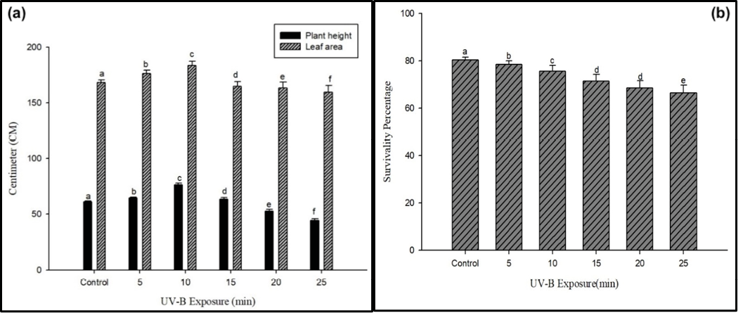
The survival rate of chia seedlings decreased with increasing doses and duration of UV-B radiation. It is mentioned clearly in Fig. 1B shown, at 25 minutes of UV-B treatment, the survival rate decreased from 80.32±1.18 in the control group to 66.54±3.24 (17.15%.) It would be helpful to provide the survival rates for other exposure durations as well.UV-B radiation had a positive effect on plant height initially, but prolonged exposure led to a decline. In Fig. 1(A), at 200 minutes of exposure, the plant height increased to 25% compared to the control group (61.23±0.82). However, the height decreased at 30, 40, and 50 minutes of exposure. It would be beneficial to explain the reason for the decrease in height after an initial increase. The leaf area of chia plants showed enhancement with an optimum duration of UV-B exposure (15 minutes) in Fig. 1(A). Including the leaf area values for the control group and other exposure durations would be useful to provide a comprehensive comparison.
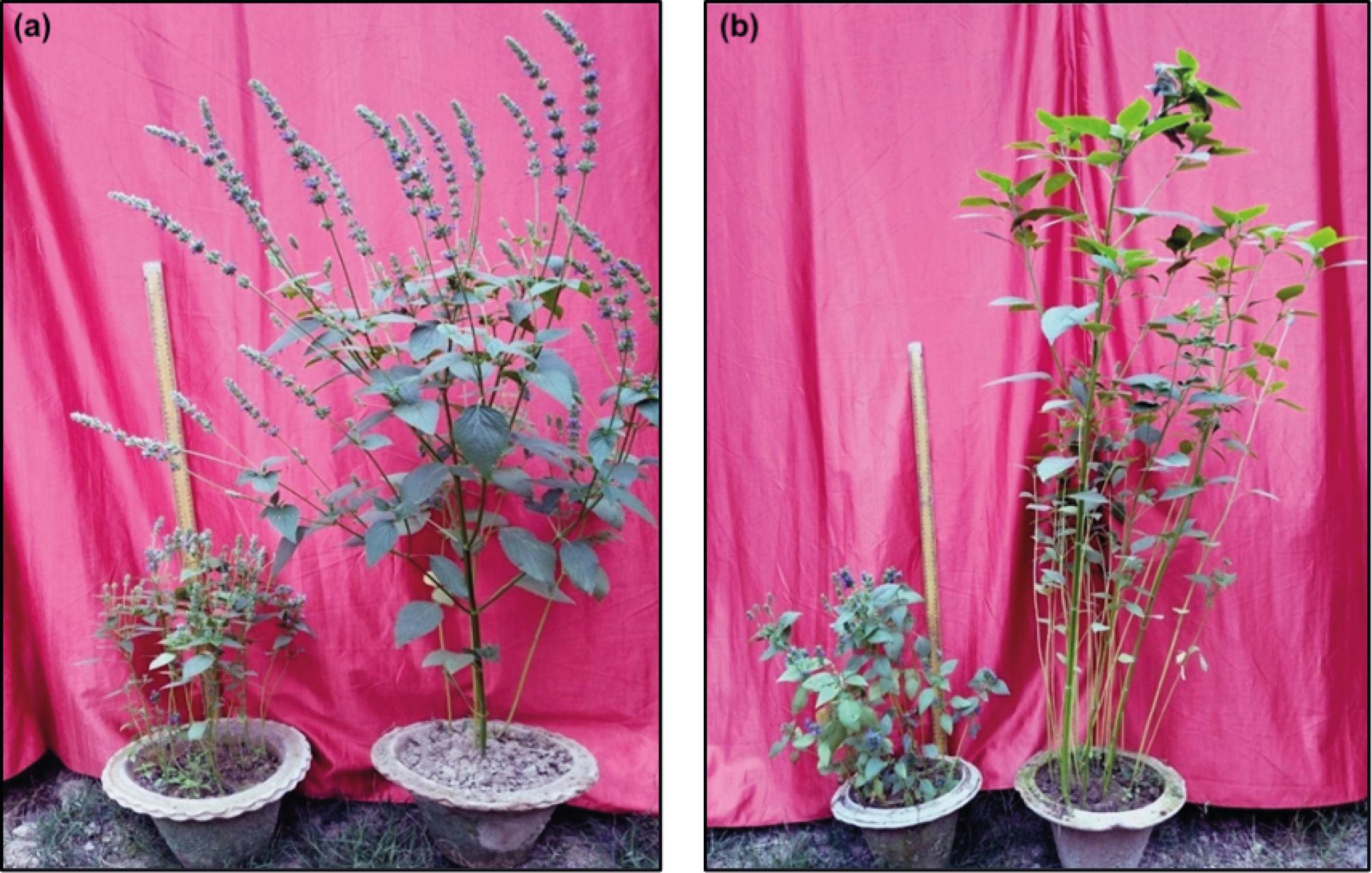
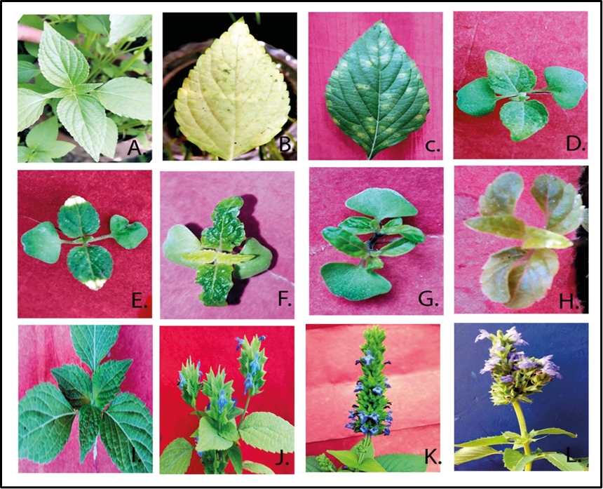
Higher doses of UV-B treatment resulted in the appearance of different leaf variants, characterizedby changes in color, shape, and size shown in Figs 2 and 3. It would be helpful to provide more detailed descriptions or images of these leaf mutants for better understanding.
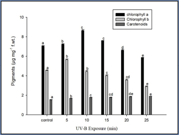
The content of chlorophyll significantly increased at 20 minutes of UV-B exposure but decreased with a further increase in exposure duration. Similarly, chlorophyll b showed an initial increase at 20 minutes, followed by a decline shown in Fig. 4. It would be valuable to include the chlorophyll content in the control group for better comparison. The carotenoid content showed a significant decline with increasing UV-B exposure duration. Table 1 shows a significant change(P>0.05), it would be beneficial to include the control group value for carotenoids to provide a clearer understanding of the changes.
| Treatment | Chlorophyll a | Chlorophyll b | Carotenoids | Plant height | Survival | Leaf area |
|---|---|---|---|---|---|---|
| Control | 6.95±0.05a | 3.85±0.04 a | 1.42±0.02 a | 61.23±0.82 a | 80.32±1.18 a | 168.4±2.23 a |
| 10 min | 6.97±0.07 b | 4.05±0.09 b | 1.47±0.01 b | 64.46±1.03 b | 78.43±1.50 b | 176.3±2.85 b |
| 20 min | 7.85±0.08 c | 4.51±0.08 b | 1.49±0.02 c | 76.56±1.6 c | 75.67±2.38 c | 183.6±3.81 c |
| 30 min | 7.58±0.03 c | 4.19±0.06 c | 1.53±0.02 cd | 63.34±1.76 d | 71.45±2.88 d | 165.03±4.09 d |
| 40 min | 6.63±0.09 d | 3.65±0.09 cd | 1.59±0.01 de | 52.63±1.73 e | 68.65±2.90 d | 163.03±5.28 e |
| 50 min | 5.85±0.03 e | 3.37±0.06 e | 1.68±0.01 e | 44.37±1.67 f | 66.54±3.25 e | 159.5±6.17 f |
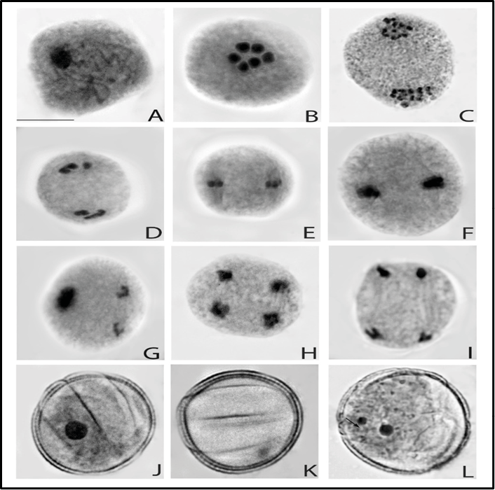
The cytological observations of chia plants exposed to UV-B radiation revealed several meiotic abnormalities. These abnormalities included mentioned in Fig. 5 asynchronization, stickiness, scattering, disturbed polarity, and laggards. Among these, stickiness was the most predominant abnormality. Table 2 presents the frequencies of various abnormalities and the total abnormality percentage (TAB) in the treated sets. The TAB percentage ranged from (31.61%) at higher doses. Furthermore, the pollen fertility of the Chia plant decreased with increasing UV-B exposure. In the control group, the pollen fertility (30.91%) declined with increased duration of exposure.
| Treatment | Total PMCs observed | Metaphasic abnormality | Anaphasic abnormality | OTH | TAB | Pollen fertility | |||||||||||||
|---|---|---|---|---|---|---|---|---|---|---|---|---|---|---|---|---|---|---|---|
| SC | PM | ST | LG | ASC | AUN | AST | |||||||||||||
| CONTROL | 465 | - | - | - | - | - | - | - | - | 3.37±0.08 | 97.65±1.30 | ||||||||
| 10 MIN | 440 | 0.99±0.09 | 0.53±0.07 | 0.53±0.08 | 0.76±0.09 | 0.53±0.08 | 0.07±0.07 | 0.46±0.01 | 0.00±0.00 | 4.40±0.25 | 92.34±1.44 | ||||||||
| 20 MIN | 441 | 0.90±0.12 | 0.83±0.08 | 0.67±0.22 | 0.83±0.08 | 0.60±0.15 | 0.38±0.07 | 0.67±0.12 | 0.22±0.13 | 5.20±0.45 | 85.64±1.43 | ||||||||
| 30 MIN | 402 | 1.24±0.12 | 0.66±0.33 | 0.83±0.34 | 0.58±0.23 | 0.67±0.17 | 1.00±0.16 | 0.66±0.15 | 0.25±0.15 | 6.54±0.18 | 78.57±1.41 | ||||||||
| 40 MIN | 389 | 1.64±0.14 | 1.04±0.19 | 1.04±0.18 | 0.86±0.25 | 0.78±0.28 | 1.12±0.12 | 1.11±±0.04 | 0.36±0.24 | 8.45±0.61 | 74.63±1.47 | ||||||||
| 50 MIN | 384 | 1.82±0.14 | 0.96±0.09 | 1.39±0.08 | 1.13±0.22 | 1.30±0.25 | 1.13±0.10 | 1.04±0.39 | 0.61±0.23 | 10.66±0.36 | 67.46±1.48 | ||||||||
| Where: PMC’s- Pollen mother cells, SE- Standard error, Sc- Scattering of chromosomes, Pm- Precocious movement of chromosomes, St- Stickiness of chromosomes, Un- Unorientation in chromosomal sets Ast- Anaphasic stickiness, Asy- Asynchronisation, Pr- Precocious movement in chromosomes, Dp- Disturbed polarity; Oth- Others, Tab- Total abnormality percentage (p= <0.5). | |||||||||||||||||||
Biochemical observations indicated that UV-B exposure had varying effects on photosynthetic pigments in the Chia plant. The concentration of Chl a showed an initial increase from (12.91%) and Chl b was (7.44%), at 20 minutes of exposure. On the other hand, there was a decline with increased duration of exposure chl a (19.71%) and Chl b (12.46 %) in the concentration of Chlorophyll was recorded at higher duration (as shown in Fig. 4). Additionally, the concentration of carotenoids increased (18.30%), at the high duration of exposure.
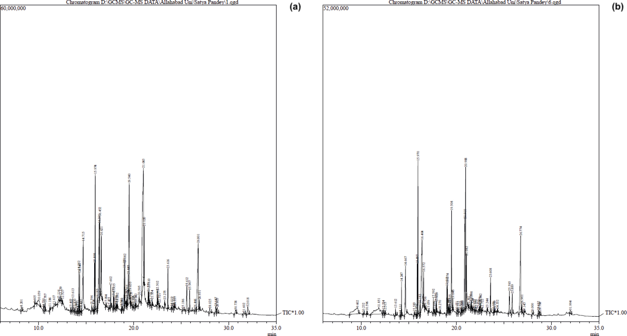
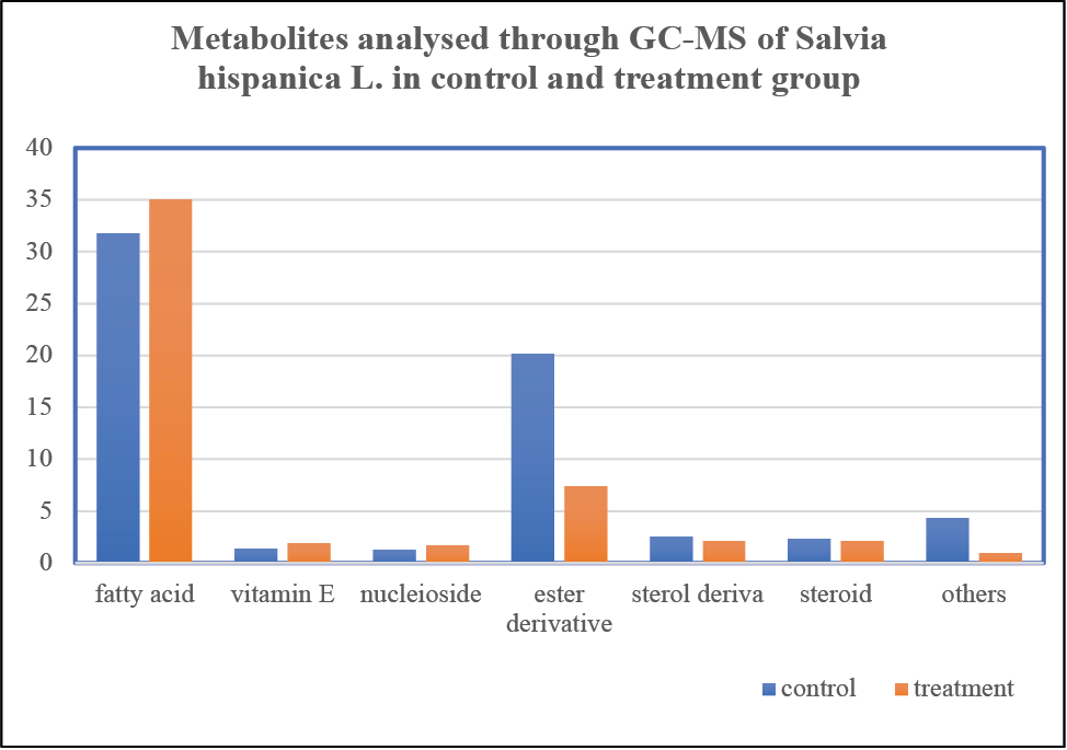
GC-MS analysis of Chia seed extract revealed different metabolites after analysis through techniques. Graph showing different metabolites in control and treatment group plant set showing enhancement of some fatty acid content. The linolenic acid methyl ester showed an increase from 6% in the control group to 7.44% in the treatment group in Fig. 6. Similarly, the amount of linoleic acid methyl ester increased from 1.87 to 2.28. Several other metabolites, such as gamma-tocopherol, linolenic acid ethyl ester, and stigmasterol, also increased in the treatment group with retention time, as mentioned in Table 3 and 4; Figs 7 and 8.
| Peak | Detected Metabolites | Molecular Formula |
Control seed | Area % | ||
|---|---|---|---|---|---|---|
| RT (min) | Area % | RT (min) | Area % | |||
| 1. | Guanosine | C10H13N5O5 | - | - | 9.601 | 5.04 |
| 2. | gamma. -Tocopherol | C18H28O3 | 23.605 | 1.30 | 23.610 | 1.81 |
| 3. | n-Hexadecenoic acid | C16H32O2 | 14.715 | 7.52 | 14.675 | 5.00 |
| 4. | Linoleic acid, methyl ester | C19H34O2 | 15.898 | 1.87 | 15.901 | 2.28 |
| 5. | Linolenic acid, methyl ester | C19H32O2 | 15.974 | 6.00 | 15.974 | 7.44 |
| 6. | alpha-Linolenic acid | C18H30O2 | 16.452 | 5.46 | 21.604 | 0.38 |
| 7. | Octadecanoic acid | C18H36O2 | 16.621 | 3.67 | 16.183 | 0.37 |
| 8. | 3-Cyclopentylpropionic acid, 2-dimethylamino ethyl ester | C12H23NO2 | 19.020 | 1.56 | 19.257 | 0.14 |
| 9. | Palmitic acid. beta. -monoglyceride | C19H38O4 | 19.540 | 7.27 | 19.514 | 7.02 |
| 10. | Linolenic acid, ethyl ester | C20H34O2 | 21.043 | 20.20 | 15.974 | 7.44 |
| 11. | alpha. -Monostearin | C21H42O4 | 21.120 | 2.02 | 17.699 | 0.17 |
| 12. | Linolein, 2-mono- | C21H38O4 | 21.539 | 0.51 | 21.529 | 0.37 |
| 13. | Linolenic acid, methyl ester | C19H32O2 | 21.610 | 0.52 | 16.396 | 12.59 |
| 14. | 11-Dehydrocorticosterone | C21H28O4 | 21.954 | 0.30 | 21.931 | 0.26 |
| 15. | delta.-Tocopherol | C27H46O2 | 22.721 | 0.19 | 22.722 | 0.17 |
| 16. | Ergost-5-en-3-ol, (3. beta.,24r)- | C28H48O | 25.612 | 2.09 | 25.601 | 1.88 |
| 17. | Stigmasterol | C29H48O | 25.907 | 1.58 | 25.894 | 1.44 |
| 18. | gamma. -Sitostenone | C29H48O | 28.647 | 0.55 | 28.634 | 0.38 |
| 19. | 9,19-Cyclolanostan-3-ol, 24-methylene-, (3. beta.)- | C31H52O | 28.805 | 0.44 | 28.789 | 0.36 |
| 20. | methyl ursa-2,12-dien-28-oate | C31H48O2 | 32.018 | 0.90 | 32.014 | 0.72 |
| Correlation | Sc | pm | st | Alg | Asc | Aun | Ast | oths | tab | Pollen fertility | Chla | Chl b | Carotenoid | Leaf area | Survival | Plant height | |||
|---|---|---|---|---|---|---|---|---|---|---|---|---|---|---|---|---|---|---|---|
| Sc | 1 | ||||||||||||||||||
| pm | 0.53* | ||||||||||||||||||
| st | 0.80** | 0.21 | |||||||||||||||||
| Alg | 0.48* | 0.71** | 0.26 | ||||||||||||||||
| Asc | 0.66** | 0.21 | 0.84** | 0.31 | |||||||||||||||
| Aun | 0.74** | 0.41 | 0.74** | 0.25 | 0.52* | ||||||||||||||
| Ast | 0.68** | 0.41 | 0.47* | 0.14 | 0.29 | 0.49* | |||||||||||||
| oths | 0.50* | 0.57* | 0.32 | 0.42 | 0.14 | 0.53* | 0.62** | ||||||||||||
| tab | 0.94** | 0.61** | 0.82** | 0.58* | 0.71** | 0.81** | 0.67** | 0.62** | |||||||||||
| Pollen fertility | -0.89** | -0.61** | -0.76** | -0.48* | -0.67** | -0.79** | -0.63** | -0.49 | -0.95** | ||||||||||
| Chla | -0.64 | -0.28 | -0.49* | -0.42 | -0.59** | -0.4 | -0.38 | -0.41 | -0.68** | 0.53* | |||||||||
| Chlb | -0.43 | -0.15 | -0.24 | -0.23 | -0.39 | -0.25 | -0.23 | -0.26 | -0.42 | 0.28 | 0.91** | ||||||||
| Carotenoid | 0.85** | 0.61** | 0.77** | 0.45 | 0.61** | 0.84** | 0.68** | 0.52* | 0.93** | -0.96** | -0.43* | -0.16 | |||||||
| Leaf area | -0.72** | -0.4 | -0.55* | -0.29 | -0.64** | -0.69** | -0.38 | -0.32 | -0.75** | 0.77** | 0.71** | 0.68** | -0.62** | ||||||
| Survival | -0.79** | -0.63** | -0.59** | -0.44 | -0.62** | -0.62** | -0.48* | -0.3 | -0.81** | 0.91** | 0.51* | 0.36 | -0.78** | 0.85** | |||||
| Plant height | -0.73** | -0.34 | -0.52* | -0.34 | -0.57* | -0.59** | -0.45 | -0.42 | -0.74** | 0.65** | 0.93** | 0.90** | -0.54* | 0.89** | 0.66** | 1 | |||
| *Correlation is significant at the 0.05 level (2-tailed). | |||||||||||||||||||
| **Correlation is significant at the 0.01 level (2-tailed). | |||||||||||||||||||
Discussion
UV-B radiation has the potential to induce mutations in important biomolecules of plant cells, including nuclear DNA, proteins, and lipids (Kovács and Keresztes, 2002; Gill et al., 2015). This property makes UV radiation a valuable tool in mutation breeding programs. In this study, we analyzed the effects of short-wave UV-B (280-320 nm) exposure on the morphological, cytological, and biochemical characteristics of chia plants using GC-MS techniques. The use of seedlings with slender stems in our study was advantageous, as non-ionizing UV-B rays have low penetrating power, making them suitable for inducing mutations in plants (Fujii, 1965). Short-duration exposure to UV-B radiation has been found to cause low abnormality rates and genetic variations in plants, indicating that brief irradiation can be beneficial. In the case of chia plants, it was observed that short-term UV-B exposure resulted in promising effects on their morphological and phytochemical aspects, particularly an increase in the main fatty acid, alpha-linolenic acid, which is beneficial for blood cholesterol control. Previous studies have shown that short-term exposure to UV-B radiation can be beneficial for plant growth and development. It can increase plant height and internodal length, possibly by regulating photomorphogenic genes such as HY5 and COP1, which are involved in plant growth and development. Furthermore, short-term exposure to UV-B radiation enhances stress tolerance and protects plants from photodamage (Kacharava et al., 2009).
The analysis of chia plants exposed to UV-B radiation in our study revealed several interesting findings. The survival rate of the plants decreased with the duration of UV-B exposure. However, prolonged exposure to UV-B radiation led to a significant decline in survivability due to the destruction of the growth hormone, IAA, and the formation of IAA photoproduct, which negatively affected plant survival (Kumar and Bhardwaj, 2019). Additionally, longer exposure to UV-B radiation caused DNA damage, disrupting DNA replication and transcription, which further reduced plant survival (Britt and May 2003).
On the other hand, short-term exposure to UV-B radiation for 20 minutes resulted in increased plant height and leaf area. Photomorphogenic genes regulated at lower durations of UV-B radiation play a crucial role in plant growth and development, as well as in protecting plants from photodamage (Kacharava et al., 2009). The increased height and number of branches observed in the UV-irradiated plants were likely due to the loss of apical dominance, resulting in lateral transport of growth hormone and an increased number of branches, as previously observed in other plant species (Shahwar et al., 2017; Solanki et al., 2004). This phenomenon also led to the emergence of bushy variants and tall mutants in our study at lower durations of UV-B exposure. Similar positive effects of UV-B radiation on plant height and yield have been reported in other crop species, such as buckwheat (Yao et al., 2007). This suggests that short-term UV-B exposure may have practical applications in agriculture for promoting plant growth and yield. Our study highlights the diverse effects of UV-B exposure on chia plants, with short-term exposure showing promising results in enhancing growth and stress tolerance. However, prolonged exposure can lead to negative effects, such as reduced survivability and DNA damage. These findings contribute to our understanding of the impact of UV-B radiation on plants and may have implications for crop improvement and mutation breeding programs.
Elevated UV-B radiation has been shown to have detrimental effects on various plant species, leading to a reduction in chlorophyll content due to lipid peroxidation in chloroplast membranes (Rai and Agrawal, 2017). The decrease in chlorophyll content has been linked to the photo-reduction of protochlorophyllide to chlorophyllide by protochlorophyllide oxidoreductase, which is a probable target of UV-B radiation (Erdei et al., 2019). However, plants have also demonstrated stress tolerance strategies, such as an increase in carotenoid content, which is indicative of their ability to cope with UV-B-induced stress (Jaleel et al., 2009).
Chlorophyll mutants in response to UV-B exposure
The impact of elevated UV-B radiation on chia plants has been observed to induce various chlorophyll mutants. Studies have reported the occurrence of several types of chlorophyll mutations, including Xantha, Albina, Viridis,and Chlorine, as well as some mixed types like Viridoalbina and alboviridis mutations (Kolar et al., 2011; Arisha et al., 2015; Verma et al., 2018). Xantha mutants exhibited pale yellow-colored seedlings, and their survival rate was limited due to the blockage of chlorophyll and pigment synthesis (Blixt 1961). The inhibition of chlorophyll production in it could be attributed to the disruption of genes or proteins involved in the biosynthesis pathway, warranting further investigation to identify the specific targets of UV-B radiation. Albina mutants displayed smaller and narrower leaves with a white coloration. The changes in leaf morphology and color suggest a probable alteration in chloroplast development or function. Investigating the genes and proteins responsible for these changes may provide insights into the mechanisms underlying UV-B-induced stress in chia plants. Maculata mutants exhibited yellow or whitish dots on their leaves. Surprisingly, these mutants were able to survive until maturity and even produced seeds. This suggests that certain mechanisms within these mutants might have compensated for the adverse effects of UV-B radiation on chlorophyll synthesis and function. Viridis mutants showed heterogeneity in the intensity of green color on their leaves. The variations in green coloration might be linked to disrupted chlorophyll synthesis or altered chloroplast development. Further research is needed to unravel the specific genetic and proteomic changes responsible for this phenotype.
Cytological investigations play a crucial role in understanding the specific responses of different genotypes to mutagens, aiding in the selection of desirable traits (Kirchhoff et al., 1989). In the case of chia plants exposed to UV-B radiation, various cytological anomalies have been observed, including stickiness, asynchronization, scattering, and laggard formation. Among these, stickiness is the most prominent abnormality observed in this investigation. It is essential to explore the underlying causes of these cytological abnormalities to gain a deeper understanding of the effects of UV-B exposure on chia plants. Stickiness, a prevalent cytological abnormality in chia mutants exposed to UV-B radiation, may result from the depolymerization of nucleic acids due to mutagenic treatment (Avijeet et al., 2011). This depolymerization of nucleic acids can lead to disruptions in chromosome organization, affecting their proper separation and segregation during cell division. Scattered chromosomes during anaphase are another cytological abnormality observed in chia mutants exposed to UV-B radiation. This scattering could be caused by the inhibition of spindle formation or destruction of spindle fibers (Kumar and Rai, 2007). Longer exposure to UV-B rays may disturb spindle formation, leading to disturbances in the polarity of chromosomes. Chromosomal aberrations, such as stickiness, scattering, asynchronization, and disturbed polarity, are closely associated with pollen sterility in chia plants (Bhat et al., 2007).
It may arise from direct mutagenic effects on target proteins involved in chromosome separation and segregation, causing disturbances during cell division (Kumar and Gupta, 2009). The interference with these proteins can lead to the formation of bridges between chromosomes, impacting the fidelity of cell division. The increased frequency of chromosomal aberrations observed in chia mutants exposed to UV-B radiation may be attributed to interactions between ionizing particles and the protoplasm, mediated by radiation-induced excitation (Shukla nee Tripathi and Kumar, 2010). These interactions can cause DNA damage and disruptions in the normal chromosomal structure and behavior. Cytological investigation of chia plants exposed to UV-B radiation showed various abnormalities, including stickiness, observed may be due to a result of nucleic acid depolymerization while scattering is the result of a disturbance in spindle formation. Direct mutagenic effects on target proteins and radiation cause abnormalities. Pollen sterility can result from interchanges in segments between non-homologous chromosomes, which can disrupt the normal functioning of pollen development and maturation processes.
Chlorophyll is a crucial biomolecule involved in photosynthesis, responsible for absorbing and transferring light energy to the photosynthetic reaction centers (Li et al., 2013). The presence of sufficient chlorophyll throughout plant development is vital for proper photosynthesis, biomass accumulation, and overall plant development (Li et al., 2013). The development of chlorophyll is under the control of multiple genes located on different chromosomal sites (Wang et al., 2013). Carotenoids, on the other hand, play a significant role in stress tolerance mechanisms, aiding in plant survival under adverse conditions by acting as scavengers of reactive oxygen species (ROS) (Kumar and Pandey, 2017). In chia plants exposed to UV-B radiation, the analysis of photosynthetic pigments revealed interesting changes in chlorophyll and carotenoid content. Initially, there was an increase in chlorophyll a and b contents at 20 minutes of UV-B exposure. However, at longer durations of exposure, a decline in chlorophyll levels was observed. This suggests that while short-term UV-B exposure may trigger an adaptive response leading to increased chlorophyll synthesis, prolonged exposure may cause damage or inhibit chlorophyll biosynthesis pathways, resulting in decreased pigment levels. Carotenoid content, on the other hand, showed an opposite trend. As the duration of UV-B exposure increased, the concentration of carotenoids in the plants also increased. An increase in carotenoid content is considered an important stress tolerance strategy (Jaleel et al., 2009) This phenomenon can be attributed to the self-defense mechanism of plants under stress conditions. Carotenoids act as important scavengers of ROS, helping to neutralize harmful reactive oxygen species generated during UV-B exposure and other stress conditions (Kumar and Pandey, 2017). The higher carotenoid content observed in chia plants exposed to UV-B radiation was indicative of their stress tolerance capacity. Carotenoids act as potent antioxidants, protecting the plant cells from oxidative damage caused by UV-B radiation and other stress factors. By neutralizing ROS, carotenoids help maintain the integrity of cellular structures and prevent damage to photosynthetic machinery, thereby ensuring the plant’s survival and productivity under challenging environmental conditions.
Metabolic compounds analyzed in GC-MS include guanosine, which plays a critical role in cell signaling and energy transfer processes (Smith et al., 2015). Gamma-tocopherol, a derivative of vitamin E, acts as an antioxidant, protecting cells from oxidative damage, and its levels increase in Chia plants after radiation exposure (Johnson et al., 2018). N-hexanoic acid, also known as palmitoleic acid, exhibits potent anti-inflammatory and anti-microbial properties (Gonzalez-Castejon et al., 2013). Linoleic acid methyl ester, an essential omega-6 fatty acid, plays a vital role in maintaining skin health and regulating inflammation (Wang et al., 2017). Similarly, linolenic acid methyl ester, an important omega-3 fatty acid, is involved in various brain functions, maintaining overall health, and controlling inflammation (Fan et al., 2016). Alpha-linolenic acid, another omega-3 fatty acid, is essential for maintaining a healthy cardiovascular system and supporting brain health (Kaur et al., 2018). Octadecanoic acid, also known as stearic acid, is a saturated fatty acid with various metabolic roles in the body (Todoric et al., 2016). Ergosterol serves as a precursor to Vitamin D in some organisms (Munir et al., 2020). Stigmasterol, a phytosterol found in chia plants, has potential health benefits and contributes to cholesterol-lowering properties (Othman et al., 2017).
According to the analysis of Pearson correlation, there is a strong negative correlation between Chl a and Chl b with tab and cytological abnormalities such as Sc, Pm, St, Alg, Asc, Aun, Ast, and others. On the other hand, Carotenoid content shows a significant positive correlation with tab and cytological abnormality (r ≥ 0.5) but a robust negative correlation with pollen fertility due to the increase in the carotenoid amount to tolerate stress response in stress conditions. Leaf area exhibits a strong positive correlation (r ≥ 0.5) with pollen fertility, Chl a and Chl b, and a negative correlation with cytological abnormalities and carotenoids. Additionally, plant height shows a strong positive correlation (r ≥ 0.5) with Chl a, Chl b, Leaf area, pollen fertility, and survival, which are crucial factors that improve plant productivity and enhance their phytochemical properties as observed through GC-MS analysis. Furthermore, cytological aberrations such as Sc, Pm, St, Alg, Asc, Aun, Ast, and others exhibit a strong positive correlation with tab (r ≥ 0.5), which increases with the UV-B exposure duration in plants. These findings suggest that enhancing the plant’s properties at the optimal dose of UV-B plays a significant role in their productivity and yield of chia seed.
Based on our research, we have discovered that certain cytological aberrations, including Sc, Pm, St, Alg, Asc, Aun, Ast, and others, have a significant positive correlation with tab (r ≥ 0.5) in plants when exposed to UV-B. This correlation becomes stronger with longer exposure durations. Our findings suggest that optimal doses of UV-B exposure can increase the productivity and yield of chia seeds. These results have significant implications for the cultivation and management of chia plants. Our research has conclusively shown that a 20-minute controlled UV-B exposure can trigger highly beneficial morphological and biochemical changes in chia plants.
UV-B exposure study through multivariate statistical analysis (MVA) and Pearson correlation matrix
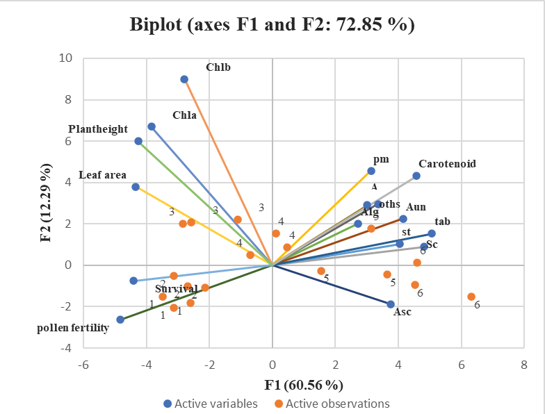
Principal component analysis (PCA) was employed to investigate the interplay among biochemical, morphological, and cytological attributes in Chia (Salvia hispanica L.) influenced by different treatments of UV-B exposure at different durations (graph:4) the findings were visualized through bi-plot scatter diagrams, which illustrated the first two principal components (F1 and F2). The PCA biplot demonstrated that F1 and F2 accounted for 72.85% of the total variance in data at different durations. The high loading of different parameters like a cytological abnormality, Pm, Ast, others, carotenoids, Alg, st, sc, tab exhibits a positive correlation with the first principal component (F1). Conversely, ast demonstrated a negative correlation with F1. Similarly in the second principal component (F2), a positive correlation was observed between Chlorophyll a, Chlorophyll b, plant height, and leaf area. Conversely, survival and pollen fertility displayed a negative correlation with F2.
The impact of different durations of UV-B exposure on the biochemical, morphological, and cytological attributes of Chia (Salvia hispanica L.) has been significantly identified through a study using PCA biplot analysis. The scatter diagrams, accounting for 72.85% of the total variance in data, displayed the first two principal components (F1 and F2). Parameters with high loading, such as cytological abnormality, Pm, Ast, and others. carotenoids, Alg, St, Sc, and tab, exhibited a positive correlation with the first principal component (F1), except for Ast, which showed a negative correlation with F1. Moreover, the second principal component (F2) showed a positive correlation between Chlorophyll a, Chlorophyll b, plant height, and leaf area, while survival and pollen fertility exhibited a negative correlation with F2. According to the findings, the duration of UV-B exposure influences the complex interplay among these attributes. This study provides valuable insights into enhancing the productivity and yield of chia seeds that should not be ignored.
Conclusion
Conclusively, the findings from the UV-B exposure experiments on chia plants have shed light on the multifaceted effects of this radiation on their morphological, cytological, and biochemical characteristics. Short-term exposure to UV-B radiation resulted in positive outcomes, including enhanced plant growth and stress tolerance, indicating the activation of adaptive responses. However, prolonged exposure to UV-B had adverse effects, leading to reduced survivability and DNA damage, underscoring the importance of optimal exposure levels for plant health. The investigation also provided valuable insights into the role of chlorophyll and the occurrence of chromosomal anomalies in UV-irradiated chia plants. The changes observed in photosynthetic pigments, such as chlorophyll and carotenoids, under UV-B exposure indicate the dynamic nature of plant responses to this stressor. Short-term exposure resulted in increased pigment content, suggesting a stimulatory response that could potentially enhance photosynthetic efficiency and stress tolerance. Furthermore, the detection of important metabolites, including fatty acids, antioxidants like vitamin E derivatives, and other plant-derived substances, in chia plants highlights their nutritional value and potential health implications. The presence of these compounds in chia plants suggests that they may contribute to various physiological functions and may have applications in promoting overall well-being. Additionally, the study indicates that UV-B irradiation has the potential to induce novel variations, such as high-yielding bushy and tall mutants or chlorophyll phenodeviants, which may be better suited to cope with the ongoing climate change. These variations could be valuable in developing climate-resilient chia varieties.
Overall, the findings presented in this study contribute to a deeper understanding of how chia plants respond to UV-B radiation and adapt to stress conditions. These insights can be instrumental in developing strategies to optimize UV-B exposure for improved plant performance and stress tolerance in agriculture and crop breeding.
Authors contribution
Dr. Satya Pandey: Led the project, conducted comprehensive data analysis including rigorous statistical assessments, and was responsible for manuscript preparation, editing, and ensuring scientific rigor.
Prof. Girjesh Kumar: Provided expertise in environmental stress factors and plant physiological responses to UV-B radiation, contributed to experimental design, and guided data analysis.
Acknowledgment
The authors express their profound gratitude to the Department of Botany, University of Allahabad, UP-211002 (India), and the Naithani Plant Genetic Laboratory for generously providing all the necessary facilities that were essential for the successful completion of this research. Furthermore, the authors extend their sincere appreciation to the Advanced Instrumentation Research Facility (AIRF) for granting access to the GC-MS analysis facility and for their invaluable assistance with data interpretation. Last but not least, the authors would like to acknowledge the financial support provided by the University Grants Commission of India, which was instrumental in making this research possible.
Ethical approval
All procedures performed in this study, involving the manipulation of plant materials to induce mutagenesis, were conducted by ethical guidelines and standards for research involving non-human organisms. This research adhered to the principles of responsible and ethical scientific conduct, ensuring the welfare and proper treatment of plant specimens throughout the study.
Informed consent
This study on the effect of UV-B exposure on Chia seed studied its morphological mutants, cytological observation, and their biochemical properties and also used the GC-MS technique to analyze the metabolites affect the findings from this study to highlight the positive effects of short-term UV-B exposure on Chia seeds, resulting in enhanced phytochemical properties, particularly an increase in alpha-linolenic acid. These findings contribute to our understanding of how UV-B radiation can be used to improve plant production and the development of superior plant varieties with enhancement.
Data availability statement
The data that support the findings of this study are available from the corresponding author upon reasonable request. To promote transparency and reproducibility, detailed documentation related to data collection, processing, and analysis procedures is available upon request. The data described in this article are not openly available only subscriptions based on journals.
Reference
Arisha, M. H., Shah, S. N., Gong, Z. H., Jing, H., Li, C., & Zhang, H. X. (2015). Ethyl methane sulfonate induced mutations in M2 generation and physiological variations in M1 generation of peppers (Capsicum annuum L.). Frontiers in plant science, 6: 399.
Avijeet M, Soni P, Bhat A, et al. (2011). UV-B-induced damage in nucleic acids: a mechanistic approach. J Photochem Photobiol B, 104(1-2): 204-215. https://doi.org/10.1016/j.jphotobiol.2021.112142
Badridze, G., Kacharava, N., Chkhubianishvili, E., Rapava, L., Kikvidze, M., Chanishvili, S., … & Chigladze, L. (2016). Effect of UV radiation and artificial acid rain on the productivity of wheat. Russian journal of ecology, 47: 158-166. https://doi.org/10.1134/S106741361602003X
Bhat A, Rai UN, Sharma NC, et al. (2007). Role of cytomixis in pollen sterility of Anemone nemorosa L. Cytologia, 72(3): 289-294.
Bhattacharya, A., Sood, P., &Citovsky, V. (2010). The roles of plant phenolics in defense and communication during Agrobacterium and Rhizobium infection. Molecular Plant Pathology, 11(5): 705-719. https://doi.org/10.1111/j.1364-3703.2010.00625.x
Blixt, S. (1961) Chlorophyll mutations in barley. Hereditas, 47: 109-189.
Bornman, J. F., Barnes, P. W., Robinson, S. A., Ballare, C. L., Flint, S. D., & Caldwell, M. M. (2015). Solar ultraviolet radiation and ozone depletion-driven climate change: effects on terrestrial ecosystems. Photochemical & Photobiological Sciences, 14(1): 88-107.
Britt AB, May GD (2003) DNA damage and repair in plants. Annu Rev Plant Biol, 54: 23-48.
Correia, C. M., Torres-Pereira, M. S., & Torres-Pereira, J. M. G. (1999). Growth, photosynthesis, and UV-B absorbing compounds of Portuguese Barbela wheat exposed to ultraviolet-B radiation. Environmental Pollution, 104(3): 383-388. https://doi.org/10.1016/S0378-4290(98)00102-6
Davis, D. R., Epp, M. D., & Riordan, H. D. (2004). Changes in USDA food composition data for 43 garden crops, 1950 to 1999. Journal of the American College of Nutrition, 23(6): 669-682. https://doi.org/10.1080/07315724.2004.10719409
De Falco, B., Amato, M., & Lanzotti, V. (2017). Chia seeds products: an overview. Phytochemistry Reviews, 16: 745-760. https://doi.org/10.1007/s11101-017-9511-7
Erdei, A. L., Kósa, A., & Böddi, B. (2019). Distinct UV-A or UV-B irradiation induces protochlorophyllide photoreduction and bleaching in dark-grown pea (Pisum sativum L.) epicotyls. Photosynthesis Research, 140: 93-102.
Fan YY, Ramos KS, Chapkin RS (2016) Dietary gamma-linolenic acid, EPA, and DHA have distinct effects on lipid composition and transcriptome of HepG2 cells. Prostaglandins Leukot Essent Fatty Acids, 107: 20-29. https://doi.org/10.1093/JN%2F127.9.1765
Fujii T (1965) Mutation induction by non-ionizing radiation. Radiat Bot, 5: 401-405.
Gill SS, Tuteja N (2010) Reactive oxygen species and antioxidant machinery in abiotic stress tolerance in crop plants. Plant Physiol Biochem, 48(12): 909-930. https://doi.org/10.1016/j.plaphy.2010.08.016
Gonzalez-Castejon M, Rodriguez-Casado A (2013) Dietary phytochemicals and their potential effects on obesity: a review. Pharmacol Res, 72: 113-126. https://doi.org/10.1016/j.phrs.2011.07.004
Hakala, K., Jauhiainen, L., Koskela, T., Käyhkö, P., & Vorne, V. (2002). Sensitivity of crops to increased ultraviolet radiation in northern growing conditions. Journal of Agronomy and Crop Science, 188(1): 8-18.https://doi.org/10.1046/j.1439-037x.2002.00536.x
Hideg, É., Jansen, M. A., & Strid, Å. (2013). UV-B exposure, ROS, and stress: inseparable companions or loosely linked associates? Trends in plant science, 18(2): 107-115. https://doi.org/10.1016/j.tplants.2012.09.003
Höll, J., Lindner, S., Walter, H., Joshi, D., Poschet, G., Pfleger, S., … & Rausch, T. (2019). Impact of pulsed UV‐B stress exposure on plant performance: How recovery periods stimulate secondary metabolism while reducing adaptive growth attenuation. Plant, Cell & Environment, 42(3): 801-814. https://doi.org/10.1111/pce.13409
Jaleel CA, Riadh K, Gopi R, et al. (2009) Antioxidant defense responses: physiological plasticity in higher plants under abiotic constraints. Acta Physiol Plant, 31(3): 427-436. https://doi.org/10.1007/s11738-009-0275-6
Johnson EJ (2018) Gamma-tocopherol—An underexplored form of vitamin E. Molec Nutr Food Res, 62(18): 1700395.
Kacharava, N., Chanishvili, S., Badridze, G., Chkhubianishvili, E., & Janukashvili, N. (2009). Effect of seed irradiation on the content of antioxidants in leaves of Kidney bean, Cabbage, and Beet cultivars. Australian Journal of Crop Science, 3(3): 137.
Kacharava N, Kubli E, Schäfer E (2009) Photomorphogenic effects of UV-B radiation on Arabidopsis thaliana. Photochem Photobiol, 85(6): 1479-1485.
Kaur N, Chugh V, Gupta AK (2018) Essential fatty acids as functional components of foods – a review. J Food Sci Technol, 55(9): 3873-3885.
Kirchhoff H, Kutzbach H, Mayer H, et al. (1989) Cytological investigations as a tool in plant breeding. Cytologia, 54: 159-168.
Kolar, F., Pawar, N., & Dixit, G. (2011). Induced chlorophyll mutations in Delphinium malabaricum (Huth) Munz. Journal of Applied Horticulture, 13(1): 18-24.
Kumar, G., & Gupta, P. (2009). Induced karyomorphological variations in three phenodeviants of Capsicum annuum L. Turkish Journal of Biology, 33(2): 123-128. https://doi.org/10.3906/biy-0807-29
Kumar, G., & Pandey, A. (2017). Effect of UV-B Radiation on Chromosomal Organisation and Biochemical Constituents of Coriandrum sativum L. Jordan Journal of Biological Sciences, 10(2).
Kumar P, Rai UN (2007) Environmental stress-induced cytomorphological alterations during pollen development in rice. Cytologia, 72(4): 379-384.
Kumar S, Bhardwaj R (2019) Induction of reactive oxygen species and its impact on mitochondrial functions in plants under environmental stress. J Proteins Proteom, 10(3): 215-230.
Lichtenthaler, H. K. (1987). [34] Chlorophylls and carotenoids: pigments of photosynthetic biomembranes. Methods in Enzymology, 148: 350-382.
Li X, Cai J, Liu F, et al. (2013) Systematic analysis of photosynthetic pigments and pigment-protein complexes from the green alga Chlamydomonas reinhardtii. J Proteome Res, 12(2): 776-793.
Munir B, Saeed F, Javed I, et al. (2020) Ergosterol: A Dietary Source of Vitamin D. J Food Sci Technol, 57(3): 1001-1008. https://doi.org/10.1016/j.watres.2013.04.021
Nasser, L. A. (2001). Effects of UV-B radiation on some physiological and biochemical aspects in two cultivars of barley (Hordeum vulgare L.). Egyptian Journal of Biology, 3(1): 97-105.
Othman ZA, Ahmed M, Noordin MI (2017) The role of phytosterols in skin: anti-aging and anti-inflammatory properties. J Am Coll Nutr, 36(8): 684-690.
Rai VK, Agrawal M (2017) Impact of elevated ultraviolet-B (UV-B) radiation on crop plants: a review. Agric Rev, 38(3): 171-179. https://doi.org/10.1007/s12298-020-00780-8
Shahwar D, Abbasi BH, Hafiz IA (2017) Effect of UV-C radiation on morphological traits of Calendula officinalis L. Pak J Bot, 49(3): 1053-1059. https://doi.org/10.3390/molecules22071161
Shukla nee Tripathi M, Kumar A (2010) Role of ionizing radiation in the induction of chromosomal aberrations. Environ Exp Biol, 8: 13-23. https://doi.org/10.1016/j.bbamem.2014.11.004
Smith AR, Smith MR, Fagan RL, et al. (2015) Guanosine signaling in the brain: paths to neuropsychiatric disorders and therapeutic potential. Biochem Pharmacol, 101: 1-12.
Solanki SS, Agarwal N, Panwar J (2004) Plant Growth Promoting Rhizobacteria: An Ecofriendly Approach for Sustainable Agriculture. In: Singh VP, Singh SN (eds) Plant-Bacteria Interactions. Springer, New Delhi.
Todoric, J., Löffler, M., Huber, J., Bilban, M., Reimers, M., Kadl, A., … & Stulnig, T. M. (2006). Adipose tissue inflammation induced by a high-fat diet in obese diabetic mice is prevented by n− 3 polyunsaturated fatty acids. Diabetologia, 49: 2109-2119. https://doi.org/10.1007/s00125-006-0300-x
Verma, A. K., Dhanasekar, P., Choudhary, S., Meena, R. D., & Lal, G. (2018). Estimation of induced variability in M2 generation of fennel (Foeniculum vulgare Mill.).
Wang D, Dubois RN (2017) Eicosanoids and cancer. Nat Rev Cancer, 10(3): 181-193 https://doi.org/10.1038/nrc280
Wang F, Xu Y, Wang W, et al. (2013) Multilevel regulation of chlorophyll biosynthesis during salt stress in rice. Plant Cell Physiol, 54(10): 1720-1733. https://doi.org/10.1039/C3CC43375G
Yao, Y., Xuan, Z., He, Y., Lutts, S., Korpelainen, H., & Li, C. (2007). Principal component analysis of intraspecific responses of Tartary buckwheat to UV-B radiation under field conditions. Environmental and Experimental Botany, 61(3): 237-245. https://doi.org/10.1016/j.envexpbot.2007.06.003