Special Issues - International Year of the Periodic Table 2019
With this Special Issues Substantia contributes to the celebrations of the 150th anniversary of the periodic table.
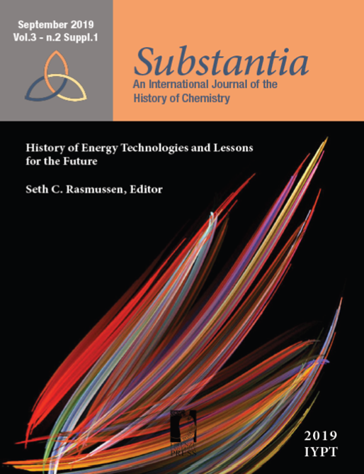 Vol 3 No 2 Suppl. 1 (2019)
"History of Energy Technologies and Lessons for the Future" edited by Seth C. Rasmussen
Cover image: Darkfield, Polarized Light micrograph (magnification 200x) from Pleurosigma (marine diatoms), by Michael J. Stringer, Westcliff-on-Sea, Essex, United Kingdom. Courtesy of Nikon Small World (1st Place, 2008 Photomicrography Competition, https://www.nikonsmallworld.com).
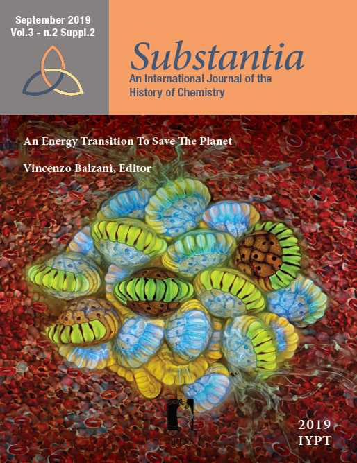 Vol 3 No 2 Suppl. 2 (2019)
"An Energy Transition To Save The Planet" edited by Vincenzo Balzani
Cover image: Autofluorescence micrograph (magnification 10x) from Fern sorus (structures producing and containing spores), by Rogelio Moreno Gill, Panama, Panama. Courtesy of Nikon Small World (2nd Place, 2018 Photomicrography Competition, https://www.nikonsmallworld.com).
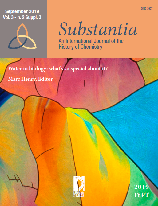 Vol 3 No 2 Suppl. 3 (2019)
"Water in Biology: What’s So Special About It?" edited by Marc Henry
Cover image: Differential Interference Contrast micrograph (magnification 40x) from a sodium vanadate crystal, by Keith Yagaloff Ph.D., Hoffmann-La Roche, Nutley, New Jersey, USA. Courtesy of Nikon Small World (8th Place, 1995 Photomicrography Competition, https://www.nikonsmallworld.com).
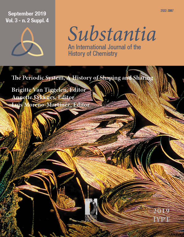 Vol 3 No 2 Suppl. 4 (2019)
"The Periodic System, a History of Shaping and Sharing" edited by Brigitte Van Tiggelen, Annette Lykknes, Luis Moreno-Martinez
Cover image: Polarized Light micrograph (magnification 45x) from scopoletin heated with chloroform and acetic acid, by Lars Bech, Naarden, The Netherlands. Courtesy of Nikon Small World (14th Place, 2000 Photomicrography Competition, https://www.nikonsmallworld.com).
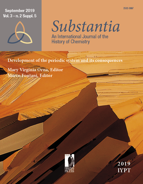 Vol 3 No 2 Suppl. 5 (2019)
"Development of the Periodic System and Its Consequences" edited by Mary Virginia Orna and Marco Fontani
Cover image: Transmitted polarized light micrograph (magnification 20x) from bisphenol-a crystallized from methanol, by John Atkinson, Union Carbide Corp., Bound Brook, New Jersey, USA. Courtesy of Nikon Small World (3rd Place, 1979 Photomicrography Competition, https://www.nikonsmallworld.com).
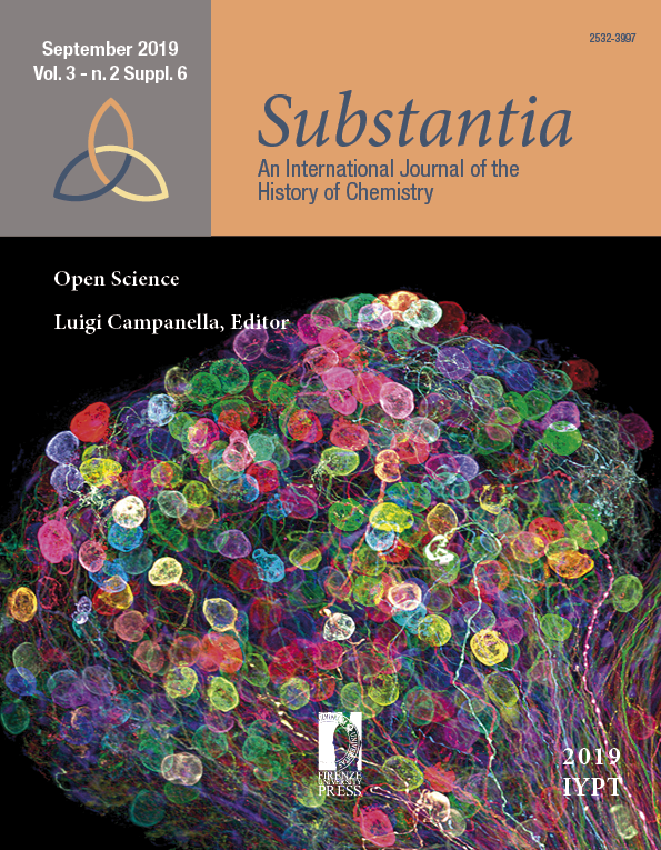 Vol 3 No 2 Suppl. 6 (2019)
"Open Science" edited by Luigi Campanella
Cover image: Confocal, tissue clearing, brainbow (labeling technique) micrograph (magnification 30x) from an individually labeled axons in an embryonic chick ciliary ganglion, by Dr. Ryo Egawa, Nagoya University, Graduate School of Medicine, Nagoya, Japan. Courtesy of Nikon Small World (7th Place, 2017 Photomicrography Competition, https://www.nikonsmallworld.com).
